

A Giant Mucinous Adenocarcinoma Arising within a Villous Adenoma of the Urachus: Case Report and Review of the Literature
- PMID: 20182635
- PMCID: PMC2825668
- DOI: 10.1155/2009/818646
A Giant Mucinous Adenocarcinoma Arising within a Villous Adenoma of the Urachus: Case Report and Review of the Literature
Abstract
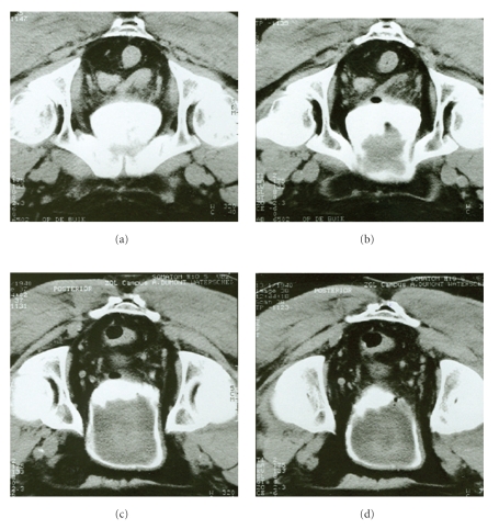
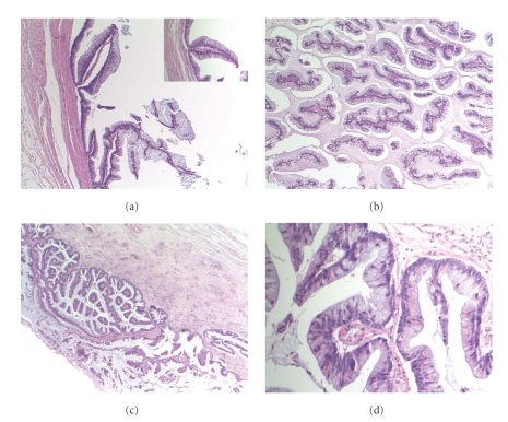
We present an exceptional case of a giant urachal tumor, consisting of both villous adenoma and mucinous adenocarcinoma of the urachus. The tumor was incidentally discovered during investigations for renal failure. Initial transurethral biopsies showed only a villous adenoma of the urachus. Although the biopsies showed no malignancy, a radical cystoprostatectomy and broad excision of the urachus and umbilicus were performed. At the same time, a bilateral nephroureterectomy was performed because of reflux-nephropathy and renal failure. The indication for surgery was based on the typical imaging aspects, raising the suspicion of an underlying urachal adenocarcinoma (size and location). Indeed, at final histopathology a concomitant well-differentiated adenocarcinoma of the urachus confined to the urachal mucosa was found. The patient remained free of disease for 50 months of follow-up. Only three previous cases of urachal adenocarcinoma associated with villous adenoma have been described.
Figures
References
-
- Sheldon CA, Clayman RV, Gonzalez R, Williams RD, Fraley EE. Malignant urachal lesions. Journal of Urology. 1984;131(1):1–8. - PubMed
-
- Mostofi MK, Thompson RV, Dean AL. Mucinous adenocarcinoma of the urinary bladder. Journal of Urology. 1955;119:68–71.
-
- Herr HW. Urachal carcinoma: the case for extended partial cystectomy. Journal of Urology. 1994;151(2):365–366. - PubMed
-
- Eble JN, Hull MT, Rowland RG, Hostetter M. Villous adenoma of the urachus with mucusuria: a light and electron microscopic study. Journal of Urology. 1986;135(6):1240–1244. - PubMed
-
- Miller DC, Gang DL, Gavris V, Alroy J, Ucci AA, Parkhurst EC. Villous adenoma of the urinary bladder: a morphologic or biologic entity? American Journal of Clinical Pathology. 1983;79(6):728–731. - PubMed
Publication types
LinkOut - more resources
Full Text Sources
