. 2010 Jul;59(7):1011-9.
doi: 10.1007/s00262-010-0823-3. Epub 2010 Feb 25.

Growth inhibition of tumor cells in vitro by using monoclonal antibodies against gonadotropin-releasing hormone receptor

Affiliations

Growth inhibition of tumor cells in vitro by using monoclonal antibodies against gonadotropin-releasing hormone receptor

Gregory Lee et al. Cancer Immunol Immunother. 2010 Jul.

Abstract

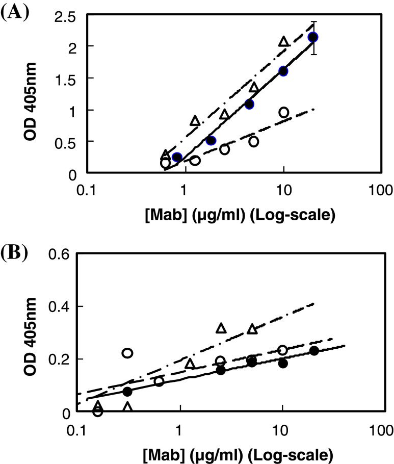
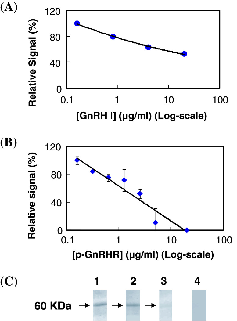
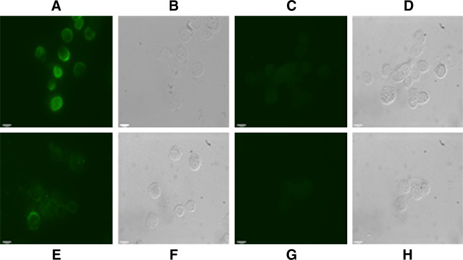
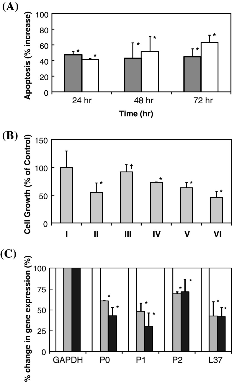
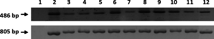
As the continuation of a previous study, synthetic peptides corresponding to the extracellular domains of human gonadotropin-releasing hormone (GnRH) receptor were used to generate additional monoclonal antibodies which were further characterized biochemically and immunologically. Among those identified to recognize GnRH receptor, monoclonal antibodies designated as GHR-103, GHR-106 and GHR-114 were found to exhibit high affinity (Kd < or = 1 x 10(-8) M) and specificity to GnRH receptor as judged by the whole cell binding immunoassay and Western blot assay. Both anti-GnRH receptor monoclonal antibodies and GnRH were shown to compete for the same binding site of GnRH receptor on the surface of cultured cancer cells. Growth inhibitions of cancer cells cultured in vitro were demonstrated by cellular apoptosis experiments (TUNEL and MTT assays) under different conditions of treatment with GHR-106 monoclonal antibody or GnRH analogs. It was generally observed that both GnRH I and GHR-106 effectively induce the apoptosis of cultured cancer cells as determined by TUNEL and MTT assays. Consistently, suppressions of gene expressions at mRNA levels were demonstrated with several ribosomal proteins (P0, P1, P2 and L37), when cancer cells were incubated with GnRH or GHR-106. The widespread expressions of GnRH receptor in almost all of the studied human cancer cell lines were also demonstrated by RT-PCR and Western blot assay, as well as indirect immunofluorescence assay with either of these monoclonal antibodies as the primary antibody. In view of the longer half life of antibodies as compared to that of GnRH or its analogs, anti-GnRH receptor monoclonal antibodies in humanized forms could function as GnRH analogs and serve as an ideal candidate of anti-cancer drugs for therapeutic treatments of various cancers in humans as well as for fertility regulations.


Figures

Fig. 1
Fig. 1
a ELISA to reveal the dose-dependent binding of GHR-103, GHR-106 and GHR-114 monoclonal antibodies to well-coated synthetic peptide corresponding to the N1-29 amino acid residues of human GnRH receptor. Following 1 h incubation at 37°C with these GHR monoclonal antibodies in microwells, goat anti-mouse IgG labeled with alkaline phosphatase was used as the secondary antibody for spectrophotometrical signal detection at 405 nm (open triangle GHR-103, filled circle GHR-106, open circle GHR-114). b ELISA to reveal the dose-dependent binding between GHR monoclonal antibodies and microwells coated with OC-3-VGH cancer cells. Experimental conditions are similar to those of Fig. 1a
Fig. 2
Fig. 2
a Inhibition of GnRH I on the binding of GHR-106 Mab (2 μg/ml) to microwells coated with OC-3-VGH cancer cells. The relative signal as percent of the maximum (in the absence of GnRH I) was plotted against GnRH I concentration (in Log Scale). b Inhibition of synthetic peptide of N1-29 amino acid residues of GnRH receptor (p-GnRHR) on the binding between GHR-106 (2 μg/ml) and microwells coated with OC-3-VGH cancer cells. The relative signal as percent of the maximum (in the absence of the peptide) was plotted against the peptide concentrations (in Log scale). The assay conditions are similar to those described in Fig. 1. c Western blot assay to reveal the protein band (s) of 60 KDa from OC-3-VGH cancer cell extract recognized by GHR-103 (Lane 1), GHR-106 (Lane 2), and GHR-114 (Lane 3). Normal mouse IgG was used as negative control (Lane 4)
Fig. 3
Fig. 3
Indirect immunofluorescence staining (under UV light) was used to demonstrate the presence of GnRH receptor on the surface of cancer cells, including OC-3-VGH ovarian cancer cell line (a) and C-33A cervical cancer cell line (e), by using GHR106 (5 μg/ml) as the primary antibody. b and f are the corresponding cells visualized under the visible light, respectively. For the negative control, the corresponding cancer cells were stained with normal mouse IgG of the same concentration as the primary antibody. The results are shown in (c, d) for OC-3-VGH and (g, h) for C-33A, respectively (magnification ×100). Details of experimental conditions were described in the text
Fig. 4
Fig. 4
a Percent increases in apoptosis in response to treatments of cultured OC-3-VGH cancer cells with 10 μg/ml of GHR-106 Mab (gray) and 0.1 μg/ml of GnRH I (white) for 24, 48 and 72 h. Percent increase in apoptosis of cancer cells was determined by subtracting spontaneous apoptosis obtained from incubation with normal mouse IgG of the same concentration or without hormone treatment for the same incubation period (all data revealed statistical significance at P < 0.05); b MTT assay to reveal dose-dependent anti-proliferative effects of I Negative control, II GnRH I (0.1 μg/ml) (P < 0.05), III Antide (0.1 μg/ml) (P > 0.05), IV Antide (0.2 μg/ml) (P < 0.05), V GHR-106 (10 μg/ml) (P < 0.05) and VI GHR-106 (20 μg/ml) Mab, respectively, for 48 h on the cultured cancer cells. Relative absorbance at 560 nm was normalized with negative control (NC) without treatment; and c down regulations of mRNA expression of four selected ribosomal proteins (P0, P1, P2 and L37) upon treatment of OC-3-VGH cancer cells with GHR-106 Mab (10 μg/ml) (gray) or GnRH I (0.1 μg/ml) (black) for an incubation period of 24 h. mRNA expression of GAPDH gene was used as the internal control and treated as 100%. Percent changes in RT-PCR signals which represent levels of P0, P1, P2, and L37 ribosomal proteins are presented with respect to GAPDH (100%) at 0 and 24 h (all data revealed statistical a significance at P < 0.05)
Fig. 5
Fig. 5
PCR products (486 bp) using GnRHR1 primers to indicate the expression of GnRHR1 chain in cancer cell lines as follows: lane 1 negative control showed no band, lane 2 Calu6, lane 3 HCT 115, lane 4 H441, lane 5 Hep3B, lane 6 HT-29, lane 7 SW-48, lane 8 SK-OV-3, lane 9 MMRU, lane 10 MRC-5, lane 11 MCF7, and lane 12 OC-3-VGH. Expression of housekeeping gene GAPDH (805 bp) is also presented to check the integrity of cDNA from each cancer cell line and to normalize the cDNA concentrations

Similar articles

Cited by

References

    1. Clayton RN. Gonadotropin-releasing hormone: its action and receptors. J Encrinol. 1989;120:11–19. doi: 10.1677/joe.0.1200011. - DOI - PubMed
    1. Lee GCY, Ho J, Chow SN, Yasojima K, Schwab C, McGeer PL. Immunoidentification of gonadotropin releasing hormone receptor in human sperm, pituitary and cancer cells. Am J Reprod Immunol. 2000;44:170–177. doi: 10.1111/j.8755-8920.2000.440307.x. - DOI - PubMed
    1. Chien CH, Chen CH, Lee CYG, Chang TC, Chen RJ, Chow SN. Detection of gonadotropin-releasing hormone receptor and its mRNA in primary human epithelial ovarian cancers. Int J Gynecol Cancer. 2004;14:451–458. doi: 10.1111/j.1048-891x.2004.014304.x. - DOI - PubMed
    1. Kang SK, Choi K-C, Yang H-S, Leung PCK. Potential role of gonadotrophin-releasing hormone (GnRH)-I and GnRH-II in the ovary and ovarian cancer. Endocr Relat Cancer. 2003;10:169–177. doi: 10.1677/erc.0.0100169. - DOI - PubMed
    1. Choi J-H, Gilks CB, Auersperg N, Leung PCK. Immunolocalization of gonadotropin-releasing hormone GnRH-I, GnRH-II, and Type I GnRH receptor during follicular development in the human ovary. J Clin Endocrinol Metab. 2006;91:4562–4570. doi: 10.1210/jc.2006-1147. - DOI - PubMed

MeSH terms