. 2009 Dec;219(11-12):577-87.
doi: 10.1007/s00427-010-0319-3. Epub 2010 Feb 25.

Partial co-option of the appendage patterning pathway in the development of abdominal appendages in the sepsid fly Themira biloba

Affiliations

Partial co-option of the appendage patterning pathway in the development of abdominal appendages in the sepsid fly Themira biloba

Julia H Bowsher et al. Dev Genes Evol. 2009 Dec.

Abstract

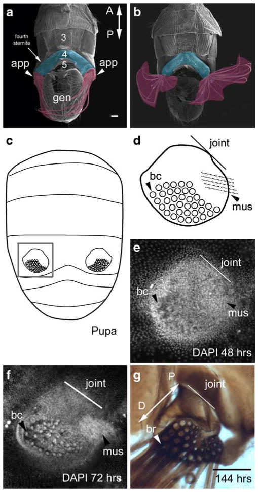
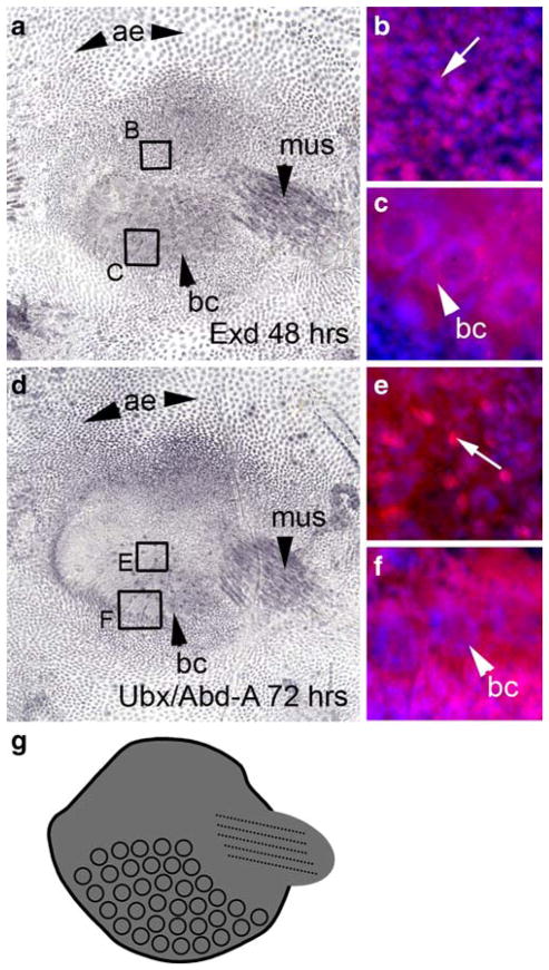
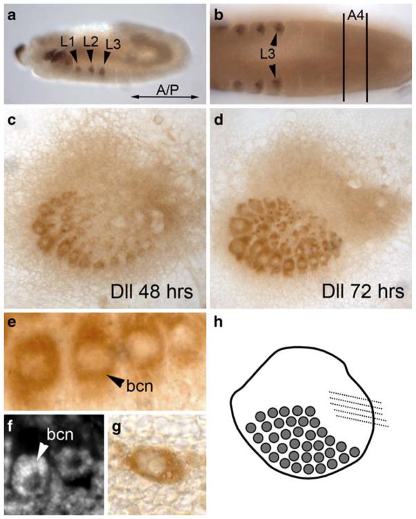
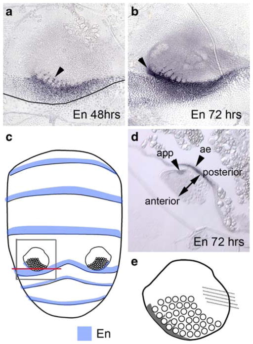
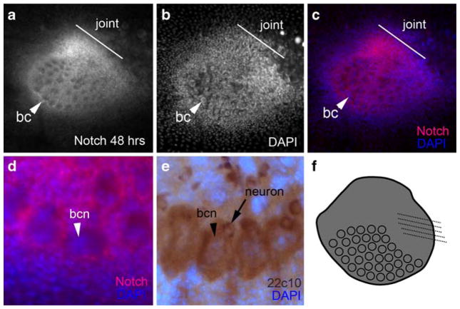
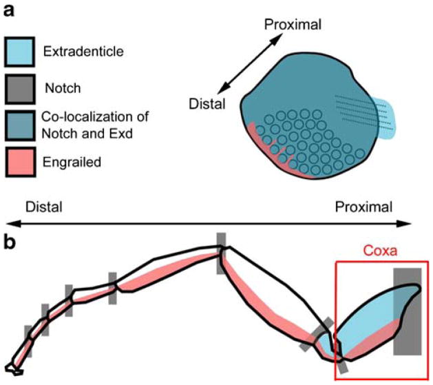
The abdominal appendages on male Themira biloba (Diptera: Sepsidae) are complex novel structures used during mating. These abdominal appendages superficially resemble the serially homologous insect appendages in that they have a joint and a short segment that can be rotated. Non-genital appendages do not occur in adult pterygote insects, so these abdominal appendages are novel structures with no obvious ancestry. We investigated whether the genes that pattern the serially homologous insect appendages have been co-opted to pattern these novel abdominal appendages. Immunohistochemistry was used to determine the expression patterns of the genes extradenticle (exd), Distal-less (Dll), engrailed (en), Notch, and the Bithorax Complex in the appendages of T. biloba during pupation. The expression patterns of Exd, En, and Notch were consistent with the hypothesis that a portion of the patterning pathway that establishes the coxopodite has been co-opted to pattern the developing abdominal appendages. However, Dll was only expressed in the bristles of the developing appendages and not the proximal-distal axis of the appendage itself. The lack of Dll expression indicates the absence of a distal domain of the appendage suggesting that sepsid abdominal appendages only use genes that normally pattern the base of segmental appendages.


Figures

Fig. 1
Fig. 1
The abdominal appendages of T. biloba are located on the fourth segment of the male abdomen. a An SEM of the ventral side of the adult male abdomen shows the paired abdominal appendages (false-colored fuschia) are attached to the lateral edge fourth sternite (false-colored blue). b These abdominal appendages have a joint and musculature, which allows for 180° rotation. c The abdominal appendages first appear during pupation. The gray box indicates the abdominal appendage shown in dg. d A cartoon of the abdominal appendage illustrates its morphological features: a large field of bristle cells (bc), a joint that connects the appendage to the body wall (line labeled ‘joint’), and a muscle (mus). e By 48 h after the beginning of pupariation, the abdominal appendage is already a distinct cluster of cells against the single-cell layer of the abdominal epidermis. The large nuclei of the bristle cells (bc) are apparent, as is the rudiment of the muscle (mus). Nuclei are stained with DAPI. f By 72 h, half-way through pupation, the morphological features or more distinct. g When the adult emerges from the pupa at 144 h, the abdominal appendage is fully formed. The long bristles (br) project toward the posterior of the abdomen. The proximal–distal axis of the appendage (double-headed arrow labeled P-D) extends from the joint where the appendage connects to the body wall (proximal) to the end of the bristle field (distal). Scale bar in a and g equals 100 μm (a adapted from Bowsher and Nijhout 2007)
Fig. 2
Fig. 2
Exd and Ubx/Abd-A are expressed throughout the appendage. a At 48 h after pupariation, Exd is expressed in the abdominal epidermis (ae), the musculature (mus) of the appendage, the developing bristle cells (bc), and the appendage itself. b A close-up of the region delineated by box B shows that Exd (magenta, Cy5) is localized to the nucleus (blue, DAPI) in the body of the appendage. The arrow indicates an individual nucleus showing Exd expression. c A close-up of the region delineated by box C shows Exd is also expressed in the bristle cells, where it is expressed in both the donut-shaped nucleus (bc) and the cytoplasm. d Expression of Ubx/Abd-A at 72 h APF appears in the abdominal appendage, the abdominal epidermis (ae), the bristle cells (bc), and the musculature (mus) of the appendage. The expression of Ubx/Abd-A coincides with that of Exd, with the exception of small regions of Ubx/Abd-A expression (arrow in e) that are not localized to a nucleus. e is a close-up of the region delineated by box E. f Ubx/Abd-A is expressed in both the donut-shaped nucleus (bc) and the cytoplasm. f is a close-up of the region delineated by box F in d. g A summary of Exd and Ubx/Abd-A expression
Fig. 3
Fig. 3
Dll is expressed in the bristle cells, but not in the rest of the abdominal appendage. a During embryogenesis, Dll expression occurs in the thoracic legs (L1, L2, and L3) of T. biloba. b During embryogenesis, there is no Dll expression in the abdomen, including the ventral side of abdominal segment four (A4), where the appendages will eventually develop. c At 48 h APF, Dll is expressed in the developing bristle cells, which has been observed in Drosophila (Campbell and Tomlinson 1998), but not in the rest of the abdominal appendage. This pattern continues at 72 h APF (d) with strong Dll expression in the bristle cells but none in the surrounding appendage. A close-up of the bristle shaft cells (e) demonstrates that the expression of Dll is localized to bristle cells only, and is not in the appendage tissue surrounding the bristles. The Dll expression in the bristle cells is nuclear (bcn). f DAPI staining of the appendage at the same stage as e shows that the bristle cell nucleus (bcn) is a donut shape. Dll expression in the bristles of the appendage is the same pattern of expression seen in other bristles cells of the abdomen, such as on the fifth abdominal segment (g). h A summary of Dll expression in the abdominal appendage
Fig. 4
Fig. 4
En is expressed in the abdominal appendage and abdominal epidermis. a En expression at 48 h APF is between the bristle cells (arrowhead) as well as in the posterior margin of the fourth segment. The boundary between the fourth and fifth abdominal segment is marked with a line. b At 72 h the appendage-specific expression is apparent in the posterior region of the appendage (arrowhead). c En (blue) is expressed in a posterior region of each abdominal segment (data not shown). The gray box delineates the region of the abdomen shown in a, b, and d. The red line indicates the location of the transverse cross-section in d. d Transverse cross-section of the appendage at 72 h shows that there are two independent regions of expression: one in the abdominal epidermis (ae) and one in the appendage (app). En expression defines the anterior–posterior axis of the appendage e a cartoon summary of En expression in the abdominal appendage
Fig. 5
Fig. 5
Notch is expressed in the abdominal appendage. a At 48 h APF, Notch is strongly expressed in the developing joint (line), the body of the appendage, and the bristle cells (bc). b A DAPI image of a. c An overlay of a and b shows the strong expression of Notch in the joint region compared to the number of nuclei. d A close-up of the bristles shows Notch expression (red) occurs in the bristle shaft cell, but not in the nucleus of that cell (bcn, blue). Expression of 22c10 (e) labels both the bristle shaft cell and the bristle neuron. f a summary of Notch expression in the abdominal appendage
Fig. 6
Fig. 6
a Gene expression in the abdominal appendage of T. biloba resembles that found at the base of the serially homologous insect appendages. b In the insect walking leg, the expression of Notch, Exd, and En all overlap in the coxa and trochanter, which are the most proximal segments of the appendage. In the abdominal appendages of T. biloba (a), Exd is expressed throughout the appendage and Dll is not expressed, indicating the absence of a distal portion

Similar articles

Cited by

References

    1. Abouheif E. Developmental genetics and homology: a hierarchical approach. TREE. 1997;12:405–408. - PubMed
    1. Abzhanov A, Kaufman TC. Homologs of Drosophila appendage genes in the patterning of arthropod limbs. Dev Biol. 2000;227:673–689. - PubMed
    1. Abu-Shaar M, Mann RS. Generation of multiple antagonistic domains along the proximodistal axis during Drosophila leg development. Development. 1998;125:3821–3830. - PubMed
    1. Angelini DR, Kauffman TC. Insect appendages and comparative ontogenetics. Dev Biol. 2005;286:57–77. - PubMed
    1. Artavanis-Tsakonas S, Matsuno K, Fortini ME. Notch signaling. Science. 1995;268:225–232. - PubMed

Publication types

MeSH terms