

Near-infrared-resonant gold/gold sulfide nanoparticles as a photothermal cancer therapeutic agent
- PMID: 20183810
- PMCID: PMC3014644
- DOI: 10.1002/smll.200901557
Near-infrared-resonant gold/gold sulfide nanoparticles as a photothermal cancer therapeutic agent
Abstract
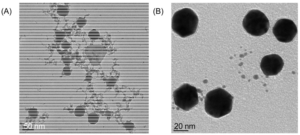
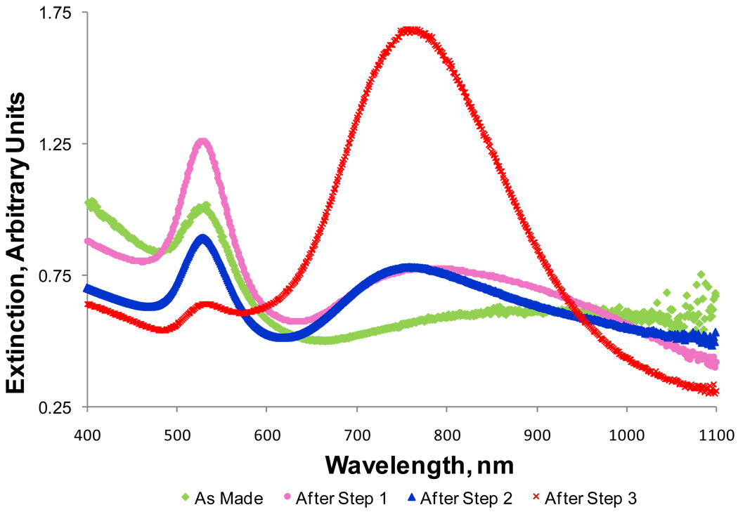
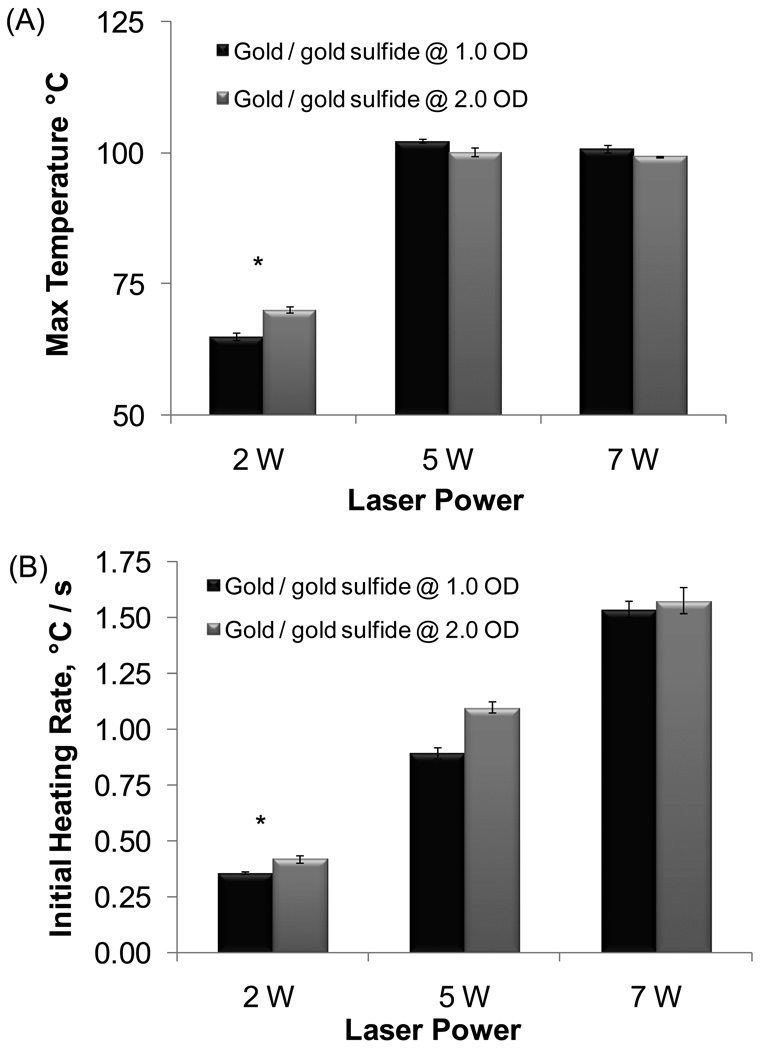
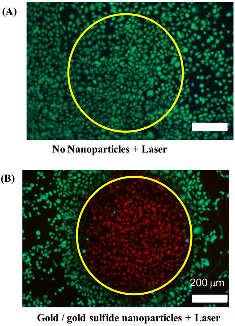
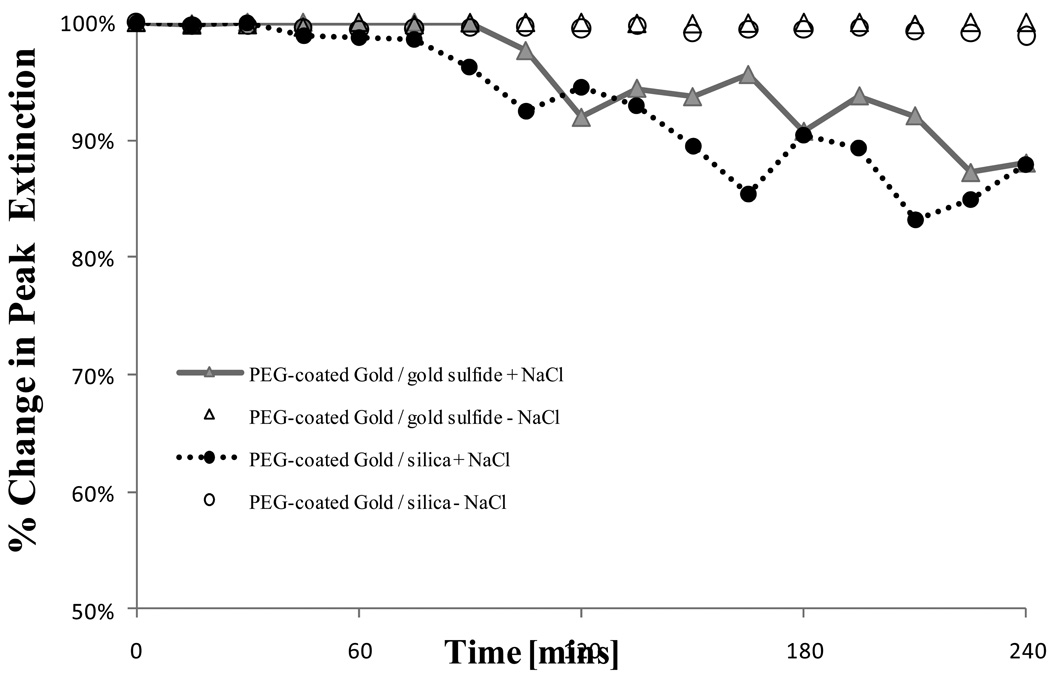
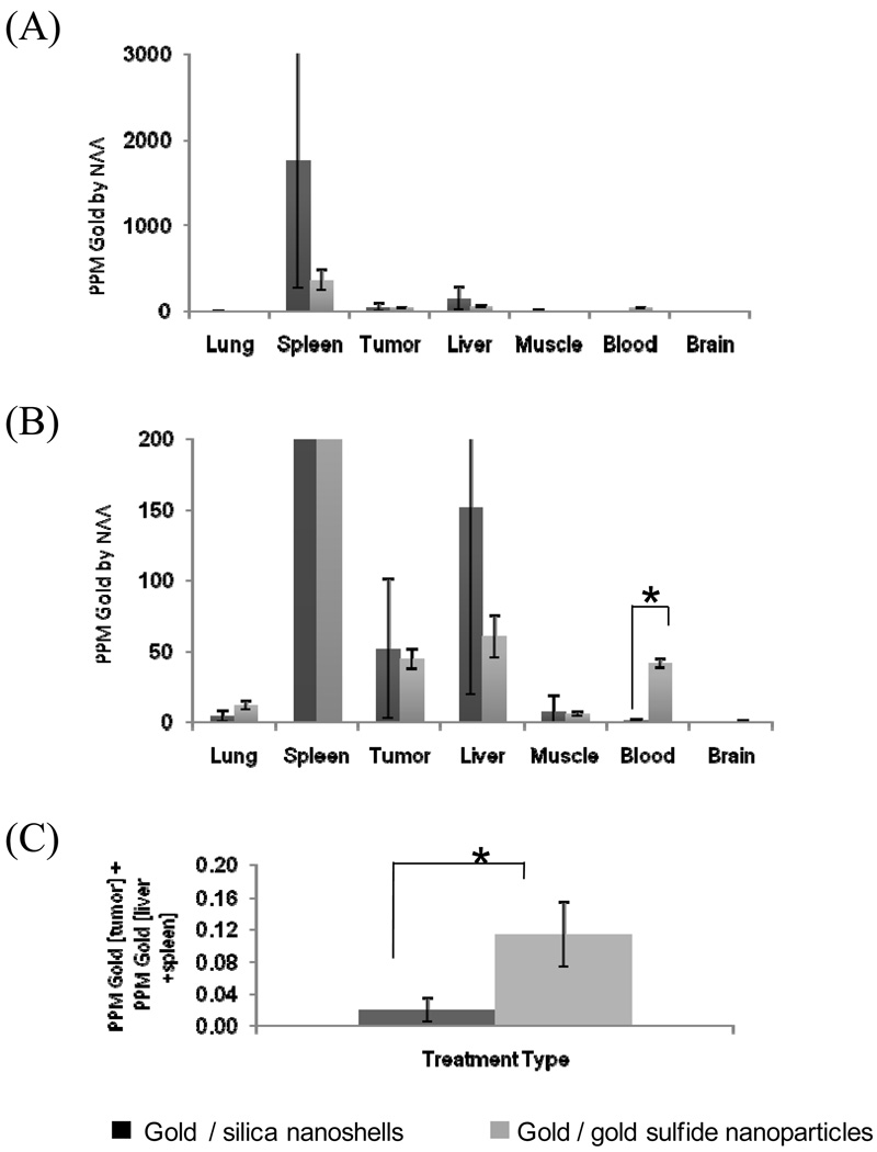
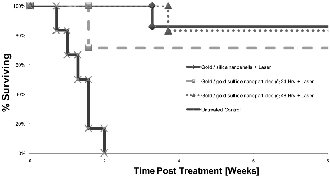
The development and optimization of near-infrared (NIR)-absorbing nanoparticles for use as photothermal cancer therapeutic agents has been ongoing. This work exploits the properties of gold/gold sulfide NIR-absorbing nanoparticles (approximately 35-55 nm) that provide higher absorption (98% absorption and 2% scattering for gold/gold sulfide versus 70% absorption and 30% scattering for gold/silica nanoshells) as well as potentially better tumor penetration. The ability to ablate tumor cells in vitro and efficacy for photothermal cancer therapy is demonstrated, and an in vivo model shows significantly increased long-term, tumor-free survival. Furthermore, enhanced circulation and biodistribution is observed in vivo. This class of NIR-absorbing nanoparticles has the potential to improve upon photothermal tumor ablation for cancer therapy.
Figures
Similar articles
-
Gold nanocages as photothermal transducers for cancer treatment.Small. 2010 Apr 9;6(7):811-7. doi: 10.1002/smll.200902216. Small. 2010. PMID: 20225187 Free PMC article.
-
Investigation of biodistribution and tissue penetration of PEGylated gold nanostars and their application for photothermal cancer treatment in tumor-bearing mice.J Mater Chem B. 2020 Jan 7;8(1):65-77. doi: 10.1039/c9tb02194a. Epub 2019 Nov 26. J Mater Chem B. 2020. PMID: 31768514
-
Contrast ultrasound-guided photothermal therapy using gold nanoshelled microcapsules in breast cancer.Eur J Radiol. 2014 Jan;83(1):117-22. doi: 10.1016/j.ejrad.2013.09.010. Epub 2013 Sep 21. Eur J Radiol. 2014. PMID: 24268740
-
Plasmonic photothermal therapy (PPTT) using gold nanoparticles.Lasers Med Sci. 2008 Jul;23(3):217-28. doi: 10.1007/s10103-007-0470-x. Epub 2007 Aug 3. Lasers Med Sci. 2008. PMID: 17674122 Review.
-
Gold nanoparticle-mediated photothermal therapy: current status and future perspective.Nanomedicine (Lond). 2014 Sep;9(13):2003-22. doi: 10.2217/nnm.14.147. Nanomedicine (Lond). 2014. PMID: 25343350 Review.
Cited by
-
Fractionated photothermal therapy in a murine tumor model: comparison with single dose.Int J Nanomedicine. 2019 Jul 18;14:5369-5379. doi: 10.2147/IJN.S205409. eCollection 2019. Int J Nanomedicine. 2019. PMID: 31409993 Free PMC article.
-
Green Synthesis of Carbon Nanoparticles (CNPs) from Biomass for Biomedical Applications.Int J Mol Sci. 2023 Jan 5;24(2):1023. doi: 10.3390/ijms24021023. Int J Mol Sci. 2023. PMID: 36674532 Free PMC article. Review.
-
Assessment of the evolution of cancer treatment therapies.Cancers (Basel). 2011 Aug 12;3(3):3279-330. doi: 10.3390/cancers3033279. Cancers (Basel). 2011. PMID: 24212956 Free PMC article.
-
Multifunctional plasmonic shell-magnetic core nanoparticles for targeted diagnostics, isolation, and photothermal destruction of tumor cells.ACS Nano. 2012 Feb 28;6(2):1065-73. doi: 10.1021/nn2045246. Epub 2012 Jan 30. ACS Nano. 2012. PMID: 22276857 Free PMC article.
-
Vascular-targeted photothermal therapy of an orthotopic murine glioma model.Nanomedicine (Lond). 2012 Aug;7(8):1133-48. doi: 10.2217/nnm.11.189. Epub 2012 May 14. Nanomedicine (Lond). 2012. PMID: 22583571 Free PMC article.
References
-
- Dalla Via L, Marciani Magno S. Curr Med Chem. 2001;8(12) - PubMed
-
- Kita K, Itoshima T, Ito T, Ogawa H, Ukida M, Kitadai M, Hattori S, Mizutani S, Tanaka R, Andoh M, et al. Gastroenterol Jpn. 1987;22(4) - PubMed
-
- Zaak D, Sroka R, Stocker S, Bise K, Lein M, Hoppner M, Frimberger D, Schneede P, Reich O, Kriegmair M, Knuchel R, Baumgartner R, Hofstetter A. Urol Int. 2004;72(3) - PubMed
-
- Loo C, Lowery A, Halas N, West J, Drezek R. Nano Lett. 2005;5(4) - PubMed
-
- Huang X, El-Sayed IH, Qian W, El-Sayed MA. J. Am. Chem. Soc. 2006;128(6) - PubMed
Publication types
MeSH terms
Substances
Grants and funding
LinkOut - more resources
Full Text Sources
Other Literature Sources
Miscellaneous
