

Review
doi: 10.1002/cphc.200900911.
Self-assembly of photosynthetic membranes
Affiliations
- PMID: 20183845
- PMCID: PMC3086839
- DOI: 10.1002/cphc.200900911
Item in Clipboard
Review
Self-assembly of photosynthetic membranes
Chemphyschem.
.
Abstract
Bacterial photosynthetic membranes, also known as chromatophores, are tightly packed with integral membrane proteins that work together to carry out photosynthesis. Chromatophores display a wide range of cellular morphologies; spherical, tubular, and lamellar chromatophores have all been observed in different bacterial species, or with different protein constituents. Through recent computational modeling and simulation, it has been demonstrated that the light-harvesting complexes abundant in chromatophores induce local membrane curvature via multiple mechanisms. These protein complexes assemble to generate a global curvature and sculpt the chromatophores into various cellular-scale architectures.
Figures
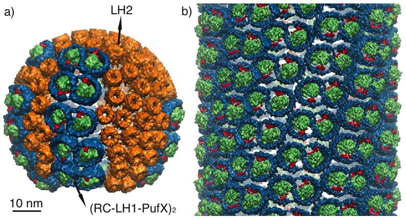
Two examples of chromatophore shapes. a) Spherical chromatophore (radius 30 nm) found in wild-type Rba. sphaeroides containing LH2 (orange) and the RC-LH1-PufX supercomplex (RC: green, LH1: blue, PufX: red). Placement of the proteins was performed in [27] using a combination of structural and imaging data. b) Mutant Rba. sphaeroides lacking LH2 possesses tubular chromatophores, which are populated with helically ordered RC-LH1-PufX.[26] The tubular chromatophore shown here has a radius of 36 nm.

a) Top view (perpendicular to membrane plane) of a setup of an LH2 simulation containing seven LH2s packed hexagonally in a membrane patch. Each LH2 is colored differently for distinction. Membrane is shown in blue, and water box in transparent blue. b) Side view (along the membrane plane) of the Rba. sphaeroides LH2 patch before and after equilibration.[28, 34] LH2s are colored according to residue type, with red being negatively-charged residues and blue positively-charged. Here, as well as in all the following protein-membrane systems shown in side views, the proteins are orientated with their cytoplasmic sides pointing upward. The dashed red lines indicate the membrane center to illustrate the curvature arising during the simulation.
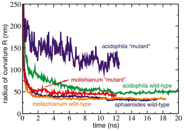
Curvature induced by patches of LH2s from wild-type vs. alanine-replacement mutants. The mutated LH2s, in which the charged residues were changed to alanines, curved less than their wild-type counterparts.
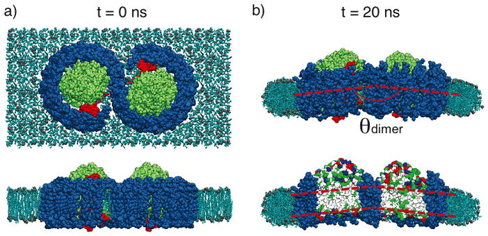
Rba. sphaeroides RC-LH1-PufX model and equilibration.[28] a) Top and side view of the RC-LH1-PufX complex immersed in a membrane patch before MD equilibration. LH1 shown in blue, RC in green, PufX in red, and membrane in light blue. Water is not shown for clarity. For the side view, some lipid molecules are hidden to show better the protein complex. An alternative placement for PufX is discussed in Scheuring et al., 2004.[37] b) Simulation system after a 20-ns equilibration. Top panel shows the complex attaining a bending angle θdimer ~ 172°. On the bottom panel, some LH1 helices are hidden to show the relative orientation of the two RCs, colored by residue types, which form a bent hydrophobic region.
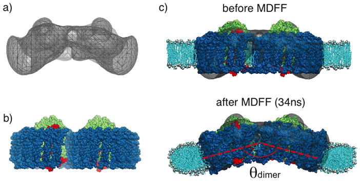
Application of MDFF for the membrane-bending properties of Rba. sphaeroides RC-LH1-PufX complex. a) EM envelope obtained in a single-molecule analysis study, showing a large bending.[26] b) Modeled RC-LH1-PufX complex from Chandler et al., 2008,[28] which is initially flat. c) RC-LH1-PufX complex fitted into the EM map employing MDFF, resulting in a much more prominent θdimer. The membrane curves along with the complex.[46]
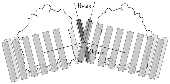
PufX dimerization and the arrangement of Rba. sphaeroides RC-LH1-PufX complex. A pair of dimerized PufX helices are shown to be located at the center of the RC-LH1-PufX complex. In this scheme, the crossing angle of the PufX helices, θPufX, leads to the bending angle of the RC-LH1-PufX, θdimer.[55] PufX shown in dark gray, LH1 in light gray, and RC in white.

Schematic diagram displaying curvature-mediated “attraction” between two membrane-bending proteins. In a), two proteins mimicking the curvature effect of the Rba. sphaeroides RC-LH1-PufX complex are placed far apart in the membrane, with each protein bending its surrounding membrane that requires energy ΔE. The shaded area in the membrane indicates the most prominent local curvature. In b), the two proteins are stacked together, and in this arrangement there is less membrane bending, indicated by fewer shaded areas. The energy required to bend the membrane is, therefore, less than the scenario depicted in a). Detailed theoretical calculations of the interactions between membrane-curving proteins can be found in several studies, e.g. Kim et al., 1998 and Chou et al., 2001.[62, 63]
References
-
- McMahon HT, Gallop JL. Membrane curvature and mechanisms of dynamic cell membrane remodeling. Nature. 2005;438:590–596. - PubMed
-
- Zimmerberg J, Kozlov MM. How proteins produce cellular membrane curvature. Nat Rev Mol Cell Biol. 2006;7:9–19. - PubMed
-
- Deserno Markus. Mesoscopic membrane physics: Concepts, simulations, and selected applications. Macromol Rapid Comm. 2009;30:752–771. - PubMed
Publication types
MeSH terms
Substances
Grants and funding
LinkOut - more resources
Full Text Sources
