Comparative Study
. 2011 Mar;15(3):635-46.
doi: 10.1111/j.1582-4934.2010.01044.x.

Tumour-associated fibroblasts and mesenchymal stem cells: more similarities than differences

Affiliations
Comparative Study

Tumour-associated fibroblasts and mesenchymal stem cells: more similarities than differences

Virgil Paunescu et al. J Cell Mol Med. 2011 Mar.

Abstract

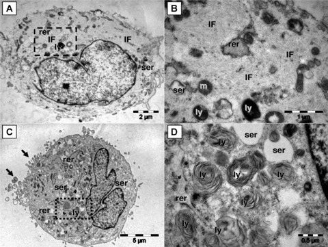
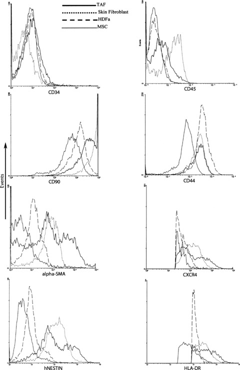
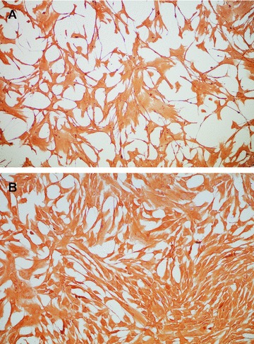
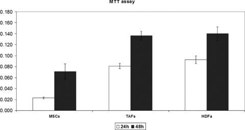
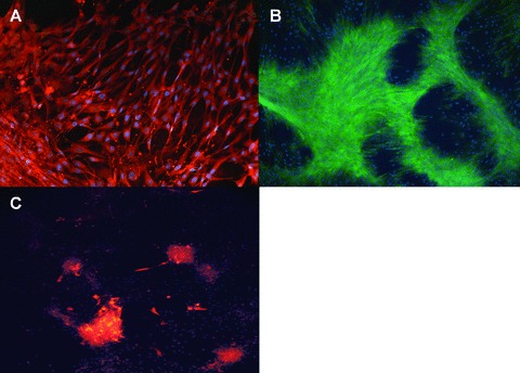
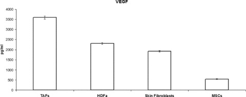
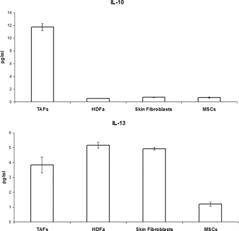
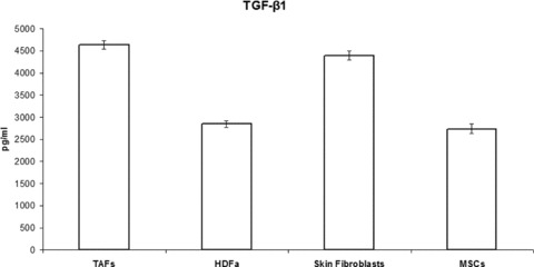
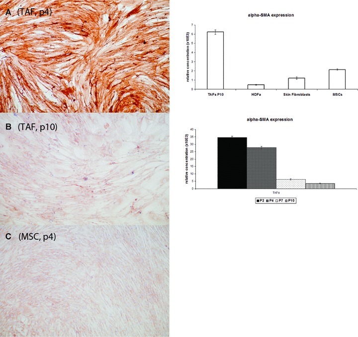
Tumour-associated fibroblasts (TAFs) are part of the tumour stroma, providing functional and structural support for tumour progression and development. The origin and biology of TAFs are poorly understood, but within the tumour environment, TAFs become activated and secrete different paracrine and autocrine factors involved in tumorigenesis. It has been shown that bone marrow mesenchymal stem cells (MSCs) can be recruited into the tumours, where they proliferate and acquire a TAF-like phenotype. We attempted to determine to what extent TAFs characteristics in vitro juxtapose to MSCs' definition, and we showed that TAFs and MSCs share immunophenotypic similarities, including the presence of certain cell surface molecules [human leukocyte antigen-DR subregion (HLA-DR), CD29, CD44, CD73, CD90, CD106 and CD117]; the expression of cytoskeleton and extracellular matrix proteins, such as vimentin, α-smooth muscle actin, nestin and trilineage differentiation potential (to adipocytes, chondrocytes and osteoblasts). When compared to MSCs, production of cytokines, chemokines and growth factors showed a significant increase in TAFs for vascular endothelial growth factor, transforming growth factor-β1, interleukins (IL-4, IL-10) and tumour necrosis factor α. Proliferation rate was highly increased in TAFs and fibroblast cell lines used in our study, compared to MSCs, whereas ultrastructural details differentiated the two cell types by the presence of cytoplasmic elongations, lamellar content lysosomes and intermediate filaments. Our results provide supportive evidence to the fact that TAFs derive from MSCs and could be a subset of 'specialized' MSCs.


Figures

Fig 1
Fig 1
Although, by light microscopy, MSCs and TAFs share a similar morphology, they exhibit major differences in their ultrastructural features. MSCs (A and B) have the cytoplasm loaded with intermediate filaments (f), few smooth (ser) and rough (rer) endoplasmic reticulum cisternae, lysosomes (ly) and mitochondria (m). Square mark in (A) is enlarged in (B). Note in (B) that intermediate filaments are organized in bundles which generate the ‘light’ appearance of the cell. The TAFs (C and D) have abundant rough endoplasmic reticulum (rer), enlarged smooth endoplasmic reticulum (ser), numerous lysosomes (ly) and grouped filopodia (arrows). Numerous lysosomes with multilamellar structure are visible in (D) [enlarged square area from (C)].
Fig 2
Fig 2
Flow cytometry analysis of cell surface markers expression on TAFs, MSCs, HDFa and skin fibroblasts. TAFs are negative for CD34 and CD45 expression but positive for all the other cell surface markers generally used to characterize MSCs. For flow cytometry cells were trypsinized and stained with specific fluorochrome-labelled monoclonal antibodies.
Fig 3
Fig 3
Vimentin expression in TAFs (A) and MSCs (B), as revealed by immunohistochemistry with a specific antibody (clone V9). Vimentin is a generic marker used to define fibroblast cells and TAFs exhibit no visible difference in the expression of this marker compared to MSCs. Magnification, 200x.
Fig 4
Fig 4
The proliferation rates of TAFs, HDFa cells and MSCs have been quantified using a standard colorimetric MTT assay that measures cellular metabolic activity. TAFs and HDFa cells were grown in culture in 96-well plates for 24 and 48 hrs and were incubated for 3 hrs with the MTT reagent at the end of the specified culture period. Both cell types show a similar proliferation rate at 24 and 48 hrs upon plating, which is higher compared to the proliferation rate of the MSCs, and in good correlation with the Trypan Blue cell viability and proliferation readings.
Fig 5
Fig 5
The trilineage differentiation potential of TAFs was assessed by immunofluorescence staining for markers specific for osteoblasts (A– osteocalcin, green fluorescence), chondroblasts (B– aggrecan, red fluorescence) and adipocytes (C– FABP4, red fluorescence). Nuclear counterstaining was performed in all cases with DAPI (blue fluorescence). Both TAFs and MSCs (photos not shown) followed a similar differentiation pattern towards osteoblasts and chondrocytes, whereas differentiation to the adipocytic lineage was 30% less efficient for TAFs compared to MSCs. Magnification, 200x.
Fig 6
Fig 6
VEGF production by TAFs, as revealed by ELISA on cell culture supernatants, is almost double compared to that of the other fibroblasts studied, and is 5-fold higher compared to that of MSCs.
Fig 7
Fig 7
Immunosuppressive cytokine secretion by MSCs and fibroblasts, as quantified by ELISA. TAFs produce, on average, 10-fold higher amounts of IL-10 compared to MSCs and the other types of fibroblasts. IL-13 secretion is similar among the different cell types.
Fig 8
Fig 8
IL-4 and TNF-α secretion by MSCs and fibroblasts measured by ELISA on cell culture supernatants at equivalent passages. TAF production of both cytokines is similar to that of the other fibroblasts and higher compared to MSCs, although the levels of IL-4 secretion were insignificantly low in all cases.
Fig 9
Fig 9
TAFs’ production of TGF-β1 is almost double compared to that of MSCs and HDFa cells, and is similar to the production of skin fibroblasts. Only the activated form of TGF-β1 was measured in our ELISA, and all the latent TGF-β1 from the supernatants was converted to the activated form prior to quantitation.
Fig 10
Fig 10
The α-smooth muscle actin (α-SMA) is highly expressed in TAFs at early passages, as revealed by immunohistochemistry (A) on cells grown in 4-well chamber slides and stained with a specific anti-human SMA antibody (clone 1A4), and by qRT-PCR with cDNA samples obtained from total RNA extracted from cells (right panel, bottom graph). However, α-SMA expression decreases significantly in the older passages (B and right panel, bottom graph). α-SMA expression is also much stronger in TAFs at early passages compared to the MSCs (C) and the other fibroblasts (qRT-PCR; right panel, upper graph).

References

    1. Friedenstein AJ, Chailakhjan RK, Lalykina KS. The development of fibroblast colonies in monolayer cultures of guinea-pig bone marrow and spleen cells. Cell Tissue Kinet. 1970;3:393–403. - PubMed
    1. Stagg J. Mesenchymal stem cells in cancer. Stem Cell Rev. 2008;4:119–24. - PubMed
    1. Liotta LA, Kohn EC. The microenvironment of the tumor-host interface. Nature. 2001;411:375–9. - PubMed
    1. Pupa SM, Menard S, Forti S, et al. New insights into the role of extracellular matrix during tumor onset and progression. J Cell Physiol. 2002;192:259–67. - PubMed
    1. Wiseman BS, Werb Z. Stromal effects on mammary gland development and breast cancer. Science. 2002;296:1046–49. - PMC - PubMed

Publication types

MeSH terms