Review
. 2010 Jun;14(6A):1199-211.
doi: 10.1111/j.1582-4934.2010.01043.x. Epub 2010 Feb 24.

Functions of ICC-like cells in the urinary tract and male genital organs

Affiliations
Review

Functions of ICC-like cells in the urinary tract and male genital organs

Hikaru Hashitani et al. J Cell Mol Med. 2010 Jun.

Abstract

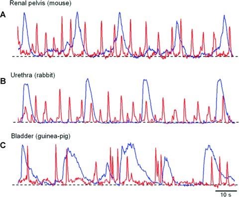
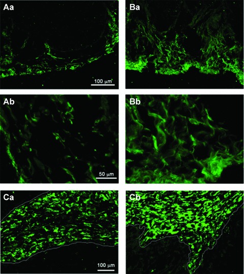
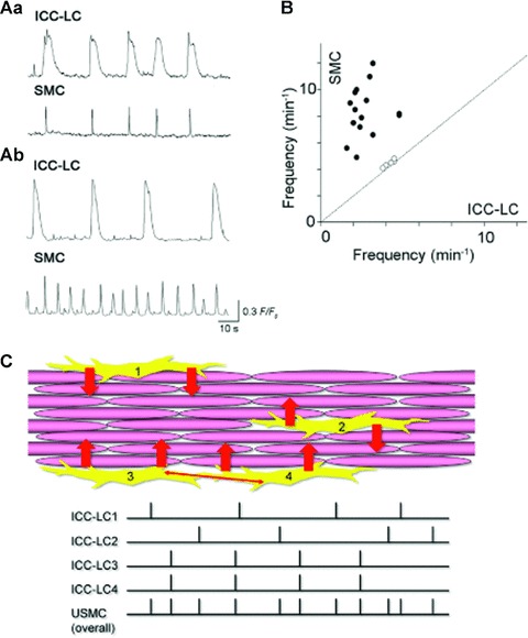
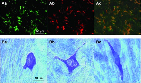
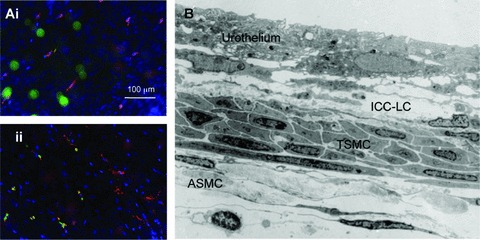
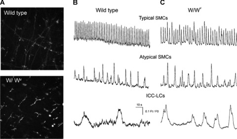
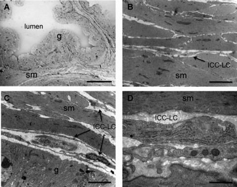
Interstitial cells of Cajal (ICC)-like cells (ICC-LCs) have been identified in many regions of the urinary tract and male genital organs by immunohistochemical studies and electron microscopy. ICC-LCs are characterized by their spontaneous electrical and Ca(2+) signalling and the cellular mechanisms of their generation have been extensively investigated. Spontaneous activity in ICC-LCs rises from the release of internally stored Ca(2+) and the opening of Ca(2+)-activated Cl(-) channels to generate spontaneous transient depolarizations (STDs) in a manner not fundamentally dependent on Ca(2+) influx through L-type voltage-dependent Ca(2+) channels. Since urogenital ICC-LCs have been identified by their immunoreactivity to Kit (CD117) antibodies, the often-used specific marker for ICC in the gastrointestinal tract, their functions have been thought likely to be similar. Thus ICC-LCs in the urogenital tract might be expected to act as either electrical pacemaker cells to drive the smooth muscle wall or as intermediaries in neuromuscular transmission. However, present knowledge of the functions of ICC-LCs suggests that their functions are not so predetermined, that their functions may be very region specific, particularly under pathological conditions. In this review, we summarize recent advances in our understanding of the location and function of ICC-LCs in various organs of the urogenital system. We also discuss several unsolved issues regarding the identification, properties and functions of ICC-LCs in various urogenital regions in health and disease.


Figures

Fig 1
Fig 1
Commonality of spontaneous Ca2+ transients recorded from ICC-LCs in the urinary tract (A) simultaneous recording of spontaneous Ca2+ transients in ICC-LCs (blue) and typical smooth muscle cells (red) of the mouse renal pelvis failed to show any temporal correlation. (B) In the rabbit urethra, the spontaneous Ca2+ transients in ICC-LCs (blue) also do not have any close temporal relationship with circular SMCs (red). (C) A simultaneous recording of spontaneous Ca2+ transients from ICC-LCs (blue) and detrusor SMCs (red) in the guinea pig bladder. Note that Ca2+ transients in ICC-LCs exhibit similar temporal characteristics among tissues as well as species.
Fig 2
Fig 2
Increased population of Kit+ and vimentin+ ICC-LCs in the obstructed bladder of the guinea pig (Aa) In normal bladder Kit-immunoreactive ICC-LCs are scattered in the subserosal and detrusor muscle layers. (Ab) Kit+ cells are spindle or stellate shaped when viewed at higher magnification. (Ba) In obstructed bladders, the number of Kit+ ICC-LCs is dramatically increased. (Bb) Kit+ ICC-LCs form close contacts with each other. (Ca) In the normal bladder vimentin+ ICC-LCs are distributed in the suburothelial layer. The white line indicates the border between the urothelium and the suburothelial layer. (Cb) In obstructed bladders the number of vimentin+ ICC-LCs is increased and their distribution expanded. Modified from [17] with permission.
Fig 3
Fig 3
ICC-LCs in the urethra act as ‘less secure’ pacemaker cells (Aa) In circular smooth muscle layer of the rabbit urethra, about 30% of ICC-LCs generated spontaneous Ca2+ transients that had a close temporal relationship with adjacent SMCs. (Ab) In the remaining 70% of cells, ICC-LCs generated spontaneous Ca2+ transients that were independent of the smooth muscle activity. (B) The frequency of ICC-LCs Ca2+ transients was invariably lower than the Ca2+ transients in the smooth muscle. (C) Model of ICC-LC pacemaking in the urethra. Individual or coupled ICC-LCs send depolarizing inputs (STDs) to adjacent SMCs. Because of low cell-to-cell electrical coupling between urethral SMCs, pacemaker signals from ICC-LCs seldom cause intercellular Ca2+ waves within muscle bundles. However, summed signal inputs from ICC-LCs to smooth muscle increase the urethras overall excitability.
Fig 4
Fig 4
COX-2 expression in Kit+ ICC-LCs and distribution of mast cells in corporal tissues (Aa, b) Scattered distribution of Kit+ ICC-LCs (Aa) and COX-2 immunoreactive cells (Ab) in rabbit corpus cavernosum. The merged picture in Ac shows Kit+ and COX-2+ cells (orange). Note that background SMCs are uniformly negative to Kit or COX-2 antibodies. (B) In the guinea pig corpus spongiosum, toluidine blue staining revealed the presence of many mast cells of various forms (round Ba, stellate Bb and spindle-shaped Bc). Modified from [45] with permission.
Fig 5
Fig 5
Presence of Kit+ cells and ICC-lCs in the mouse renal pelvis (A) fluorescence micrographs of the lamina propria (Ai) and serosal (Aii) regions of an unfixed whole mount preparation of mouse renal pelvis exposed to FITC FD70S (green) to label macrophages and then the extraceullarly binding Kit antibody H300 (red) and followed by the nuclear stain, Hoechest blue. Less than 20% of Kit+ cells also displayed the FITC-dextran fluorescence. (B) Low magnification electron micrograph through the mid region of the mouse renal pelvis illustrating the urothelium, ICC-LCs within the lamina propria and both typical (TSMC) and atypical (ASMC) SMCs.
Fig 6
Fig 6
Comparison of Kit+ cells and Ca2+ signalling in the renal pelvis of wild-type and W//Wv mice (A) fluorescence micrographs of unfixed whole mount preparations of mouse renal pelvis obtained from wild-type and W/Wv transgenic mice and exposed to the Kit antibody AK2. Ca2+ waves in typical SMCs in the absence of nifedipine and Ca2+ transients recorded in atypical SMCs and ICC-LCs recorded in the presence of 1 μM nifedipine were indistinguishable in wild-type (B) and W/Wv mice (C).
Fig 7
Fig 7
ICC-like cells in the guinea pig prostate (A) Low magnification light micrograph of the junction between 3 prostatic acini within the ventral lobe of the guinea pig prostate illustrating that the lumen is surrounded by a glandular secretory epithelial cells (g) enveloped by a stroma consisting mainly of SMCs (sm). Scale bar represents 50 μm. Higher magnification electron microscopy demonstrates that the stroma is tightly packed with SMCs (B) and that ICC-LC cells are found within the space between the lamina propria and the epithelial cells (C). ICC-LC processes were also found between the SMCs (B); scale bars represent 2 μm. (D) higher magnification of an ICC-like cell illustrating internal organelles such as the rough endoplasmic reticulum and cavaeloe, scale bar 0.5 μm.

Similar articles

Cited by

References

    1. Sanders KM, Koh SD, Ward SM. Interstitial cells of Cajal as pacemakers in the gastrointestinal tract. Annu Rev Physiol. 2006;68:307–43. - PubMed
    1. Brading AF, McCloskey KD. Mechanisms of disease: specialized interstitial cells of the urinary tract–an assessment of current knowledge. Nat Clin Pract Urol. 2005;2:546–54. - PubMed
    1. McCloskey KD, Anderson UA, Davidson RA, et al. Comparison of mechanical and electrical activity and interstitial cells of Cajal in urinary bladders from wild-type and W/Wv mice. Br J Pharmacol. 2009;156:273–83. - PMC - PubMed
    1. Johnston L, Sergeant GP, Hollywood MA, et al. Calcium oscillations in interstitial cells of the rabbit urethra. J Physiol. 2005;565:449–61. - PMC - PubMed
    1. Hashitani H, Suzuki H. Properties of spontaneous Ca2+ transients recorded from interstitial cells of Cajal-like cells of the rabbit urethra in situ. J Physiol. 2007;583:505–19. - PMC - PubMed

Publication types