

Progressive ossification due to retained surgical sponge after upper leg amputation: a case report
- PMID: 20184688
- PMCID: PMC2827071
- DOI: 10.1186/1757-1626-0002-0000008592
Progressive ossification due to retained surgical sponge after upper leg amputation: a case report
Abstract
Introduction: Numerous cases are described of patients in whom foreign objects were found after surgery. Foreign body granuloma caused by retained surgical sponge, also called gossypiboma, mostly occur in the abdominal cavity but very seldom in limbs.
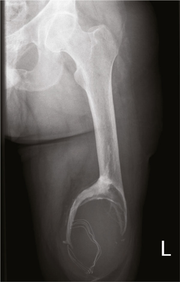
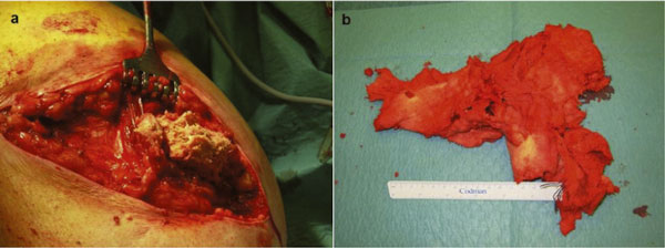
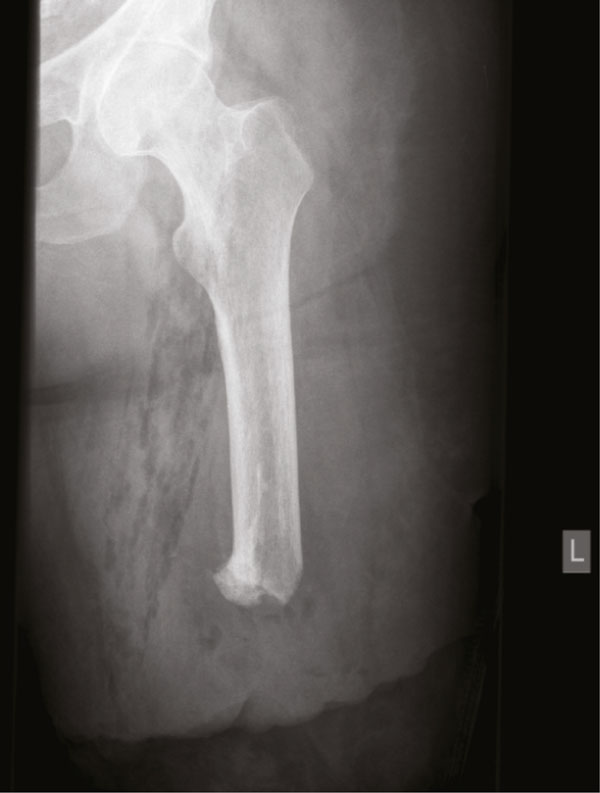
Case presentation: A 29-year-old Caucasian man presented with asymmetrical walking pattern and progressive pain in his leg, which was severely injured and amputated seven years before. A firm swelling of soft tissue with calcifications was localized in the stump. Roentgenogram and MRI showed a retained surgical sponge with calcifications. Open surgery was performed and a well-encapsulated, brownish soft-tissue tumour containing serous fluid was found in which the remnants of a surgical sponge of 40 x 40 centimeters was identified and removed. Infectious complications characterized the postoperative course for which multiple surgical procedures were needed to create a definitive healing of the stump.
Conclusion: A surgical sponge left behind in an amputated leg may lead to fibroma, destruction, osteolysis and calcification. In our case the gauze lead to mild dysfunction of the prosthetic leg, asymmetrical walking pattern, phantom pain and calcification and osteolysis on roentgenogram.
Figures
References
LinkOut - more resources
Full Text Sources
