

Benign splenosis mimicking peritoneal seeding in a bladder cancer patient: a case report
- PMID: 20184706
- PMCID: PMC2827057
- DOI: 10.1186/1757-1626-0002-0000008982
Benign splenosis mimicking peritoneal seeding in a bladder cancer patient: a case report
Abstract
Introduction: Splenosis is a post-traumatic autotrasplantation and proliferation of splenic tissue in ectopic sites. These implants may mimic malignancy in healthy patients or peritoneal metastases in cancer patients. When a previous history of splenic injury is known, the finding of soft tissue nodules in many thoracic and abdominal locations might raise the suspicion of the benign condition of splenosis, in order to avoid unnecessary surgery or chemotherapy.
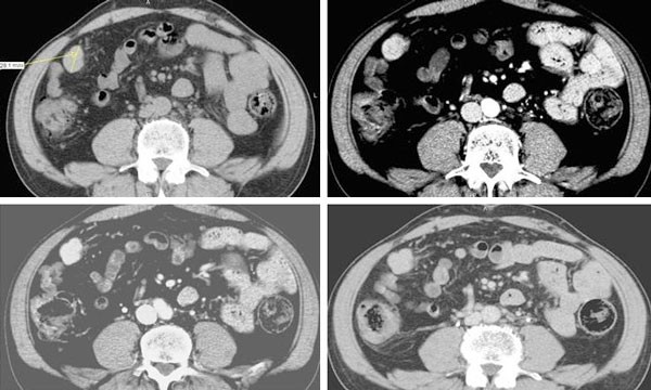
Case presentation: A 56-year-old man with history of persistent hematuria from bladder cancer was referred to our Institution for suspected peritoneal carcinosis. For staging purposes he underwent abdominal computed tomography and ultrasound. The integration of patient's history and imaging results led to the diagnosis of peritoneal splenosis. The patient therefore underwent regular Trans Urethral Resection of Bladder for the known malignancy; while no treatment was necessary for splenosis. Two years follow-up was negative for metastases.
Conclusion: Splenosis is a benign condition after traumatic splenectomy which should be taken into account in the differential diagnosis with peritoneal seeding of malignancy because its appearance may resemble malignancy.
Figures

References
-
- Khosravi MR, Margulies DR, Alsabeh R, Nissen N, Phillips EH, Morgenstern L. Consider the diagnosis of splenosis for soft tissue masses long after any splenic injury. Am Surg. 2004;70:967–970. - PubMed
-
- Lin WC, Lee RC, Chiang JH, Wei CJ, Chu LS, Liu RS, Chang CY. MR features of abdominal splenosis. AJR Am J Roentgenol. 2003;180:493–496. - PubMed
-
- Normand JP, Rioux M, Dumont M, Bouchard G, Letourneau L. Thoracic splenosis after blunt trauma: frequency and imaging findings. AJR Am J Roentgenol. 1993;161:739–741. - PubMed
LinkOut - more resources
Full Text Sources
