. 2010 Feb 25:5:6.
doi: 10.1186/1749-8104-5-6.

Nav2 is necessary for cranial nerve development and blood pressure regulation

Affiliations

Nav2 is necessary for cranial nerve development and blood pressure regulation

Elizabeth M McNeill et al. Neural Dev. .

Abstract

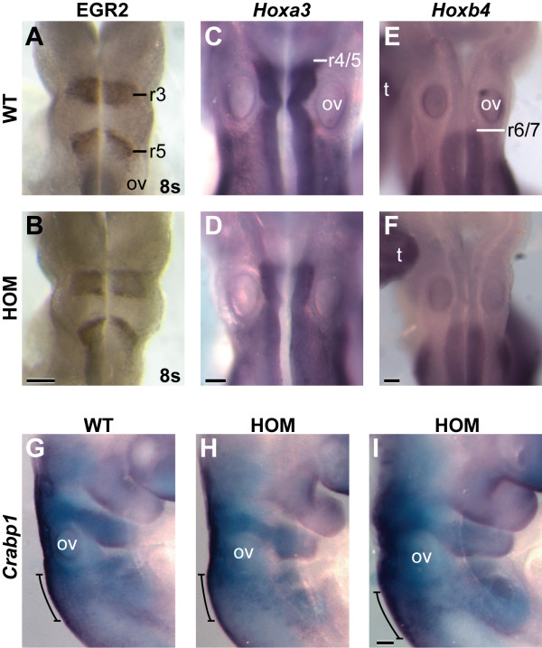
Background: All-trans retinoic acid (atRA) is required for nervous system development, including the developing hindbrain region. Neuron navigator 2 (Nav2) was first identified as an atRA-responsive gene in human neuroblastoma cells (retinoic acid-induced in neuroblastoma 1, Rainb1), and is required for atRA-mediated neurite outgrowth. In this paper, we explore the importance of Nav2 in nervous system development and function in vivo.

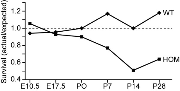
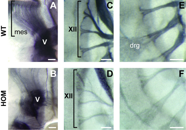
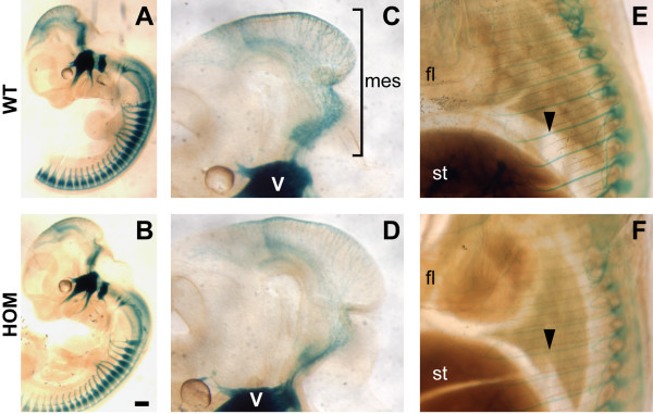
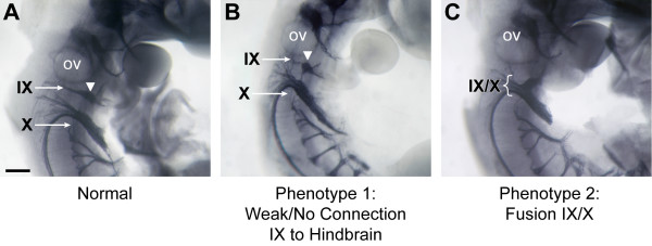
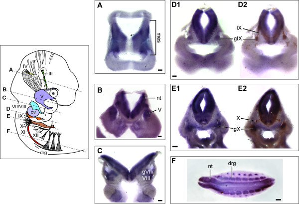
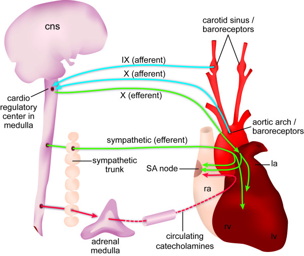
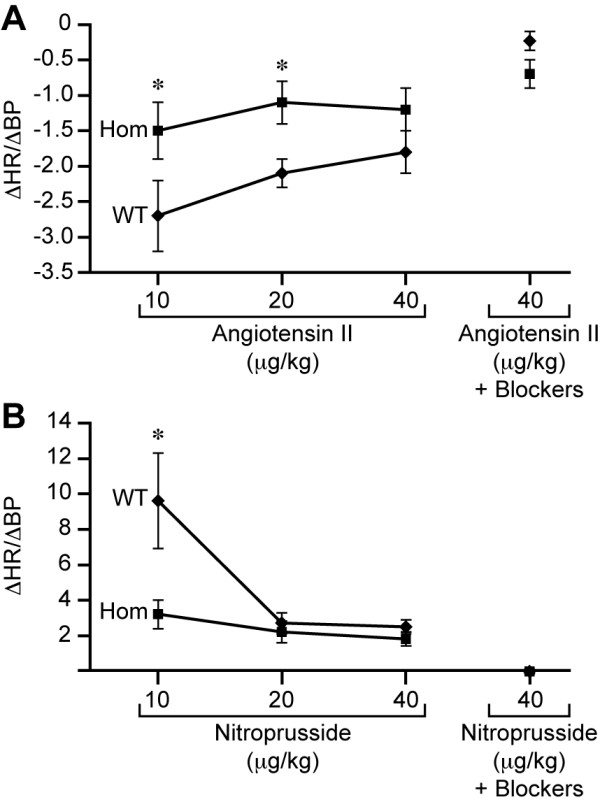
Results: Nav2 hypomorphic homozygous mutants show decreased survival starting at birth. Nav2 mutant embryos show an overall reduction in nerve fiber density, as well as specific defects in cranial nerves IX (glossopharyngeal) and X (vagus). Nav2 hypomorphic mutant adult mice also display a blunted baroreceptor response compared to wild-type controls.

Conclusions: Nav2 functions in mammalian nervous system development, and is required for normal cranial nerve development and blood pressure regulation in the adult.


Figures

Figure 1
Figure 1
Postnatal survival is reduced in Nav2-/- mice. Female Nav2 heterozygotes were mated with Nav2+/- and Nav2-/- males, and litters were examined and genotyped at embryonic days (E) 10.5 and 17.5, and postnatal days (P) 0, 7, 14, and 28; with a total of 90, 188, 348, 94, 93, and 791 offspring examined at each time point, respectively. A reduction in expected number live animals (<1.0) appears in the homozygote mutant (HOM) group after P0. At weaning (P28), only 60% of the expected number of homozygous mice are alive. Wild-type (WT) values remain around the expected ratio of 1.0 (indicated by the dotted line) at all time points.
Figure 2
Figure 2
Nerves are less densely stained by an antibody to neurofilament in Nav2 homozygous mutant embryos at E11.5. The (A) mesencephalic tract (mes) nerve fibers, (C) cranial nerve XII, and (E) dorsal root ganglia (drg) nerves are shown in wild-type (WT) mice. (B, D, F) Nav2-/- littermates show decreased staining of nerve fibers in these same regions. The images shown here are representative of the majority of those scored in Table 1 as positive for a reduction of nerve fiber density. Scale bar: 100 μm.
Figure 3
Figure 3
Nav2-/-/Brn3a-lacZ+/- mice show a reduction in lacZ staining of sensory nerves compared to their Nav2+/+/Brn3a-lacZ+/- littermates. (A) Nav2 wild-type (WT) and (B) homozygote mutant (HOM) littermates in a Brn3a-lacZ+/- background shown at the same developmental stage (E13.5). (C, D) Specifically, there appear to be fewer sensory neurons in the mesencephalic tract (mes) region in the Nav2-/-/Brn3a-lacZ+/- embryo (D) (E13.5) compared to the wild-type (WT) littermate (C) (E13.5). (E, F) A reduction in spinal nerve staining is also observed in the Nav2-/-/Brn3a-lacZ+/- embryo at E15.5 (compare (F) to the wild-type in (E)). The results shown are representative of nine wild-type and nine Nav2-/- embryos examined at E12.5 to 13.5, and two wild-type and three Nav2-/- embryos at E15.5. Scale bar: 200 μm. fl, forelimb; st, stomach.
Figure 4
Figure 4
Cranial nerve IX and X defects are found in homozygous Nav2 hypomorphic mutants. Whole-mount immunostaining for neurofilament protein in (A) a normal Nav2+/+ embryo, and in (B, C) Nav2-/- embryos at E11.5. In the Nav2-/- embryo with phenotype 1 (B), the nerve fibers connecting the distal ganglia of IX (white arrowhead) to the proximal ganglia and hindbrain exit point are missing. In the Nav2 mutant showing phenotype 2 (C), the IXth and Xth nerves are fused (brackets). Cranial nerves are indicated in roman numerals: IX, glossopharyngeal; X, vagus; ov, otic vesicle. Scale bar: 200 μm.
Figure 5
Figure 5
Nav2 is expressed in the mesencephalic tract, cranial and dorsal root ganglia and neural tube at E10.5. (A-F) Nav2 mRNA expression in transverse vibratome sections of a wild-type embryo. Representative sections (200 μm) were analyzed by in situ hybridization using a riboprobe to Nav2. Staining for Nav2 is present in the mesencephalic tract (mes) neurons (A), trigeminal ganglia (V) (B), ganglia of VII and VIII (C), the ganglia of IX (D1) and X (E1) as well as the dorsal root ganglia (drg) (F). β-III tubulin antibody staining in vibratome sections (D2, E2) overlaps with that of the Nav2 riboprobe in the ganglia of cranial nerve IX (D1) and X (E1). The approximate location of the vibratome sections with respect to the whole embryo is shown by a dotted line in the illustration. e, eye; nt, neural tube; ov, otic vesicle. Scale bar: 200 μm.
Figure 6
Figure 6
Expression of EGR2 (KROX20), Hoxa3, Hoxb4, and Crabp1 is unaltered in the hindbrain of Nav2 homozygous mutant embryos. Whole-mount immunohistochemistry for EGR2 shows staining in presumptive rhombomeres r3 and r5 at 8s in both (A) wild-type (WT) and (B) Nav2-/- (HOM) embryos. (C,D) In situ hybridization analysis of Hoxa3 mRNA at E9.5 shows staining in the neuroepithelium caudal to the r4/5 border in both genotypes. Hoxb4 mRNA at E9.5 is expressed caudal to the r6/7 border in both (E) wild-type and (F) Nav2-/- embryos. (G-I) Crabp1 mRNA at E9.5 is expressed in the neural crest cells, including those contributing to the IXth and X cranial nerves (bracket) in both wild-type (G) and Nav2-/- (H-I) embryos. A total of ten 8s wild-type and Nav2-/- embryos each were studied for EGR2 staining; 8 and 19, and 7 and 12 wild-type and Nav2-/- embryos were examined for Hoxa3 and Hoxb4, respectively. Seven wild-type and 13 Nav2-/- embryos were studied for Crabp1 staining. ov, otic vesicle; t, tail. Scale bar: 200 μm.
Figure 7
Figure 7
Diagram of sympathetic and parasympathetic regulation of the baroreceptor reflex. Schematic shows the nerves involved and areas innervated in the baroreceptor reflex arc. Adapted from [63]. CNS, central nervous system; la, left atrium; lv, left ventricle; ra, right atrium; rv, right ventricle; SA, sinuatrial.
Figure 8
Figure 8
The baroreceptor reflex response in response to angiotensin II and nitroprusside. Nav2 hypomorphic mutants (Hom) show blunted responsivity (change in heart rate (ΔHR)/change in blood pressure (ΔBP)) at varying doses of (A) angiotensin II and (B) nitroprusside. Asterisks indicate statistical significance at P < 0.001 as determined by a two tailed t-test. WT, wild-type.

Similar articles

Cited by

References

    1. Gavalas A, Krumlauf R. Retinoid signalling and hindbrain patterning. Curr Opin Genet Dev. 2000;10:380–386. doi: 10.1016/S0959-437X(00)00100-3. - DOI - PubMed
    1. Clagett-Dame M, DeLuca HF. The role of vitamin A in mammalian reproduction and embryonic development. Annu Rev Nutr. 2002;22:347–381. doi: 10.1146/annurev.nutr.22.010402.102745E. - DOI - PubMed
    1. Wilson L, Maden M. The mechanisms of dorsoventral patterning in the vertebrate neural tube. Dev Biol. 2005;282:1–13. doi: 10.1016/j.ydbio.2005.02.027. - DOI - PubMed
    1. Glover JC, Renaud JS, Rijli FM. Retinoic acid and hindbrain patterning. J Neurobiol. 2006;66:705–725. doi: 10.1002/neu.20272. - DOI - PubMed
    1. White RJ, Schilling TF. How degrading: Cyp26s in hindbrain development. Dev Dyn. 2008;237:2775–2790. doi: 10.1002/dvdy.21695. - DOI - PMC - PubMed

Publication types

MeSH terms