. 2010 Feb 25:10:64.
doi: 10.1186/1471-2180-10-64.

Modulation of iron homeostasis in macrophages by bacterial intracellular pathogens

Affiliations

Modulation of iron homeostasis in macrophages by bacterial intracellular pathogens

Xin Pan et al. BMC Microbiol. .

Abstract

Background: Intracellular bacterial pathogens depend on acquisition of iron for their success as pathogens. The host cell requires iron as an essential component for cellular functions that include innate immune defense mechanisms. The transferrin receptor TfR1 plays an important part for delivering iron to the host cell during infection. Its expression can be modulated by infection, but its essentiality for bacterial intracellular survival has not been directly investigated.

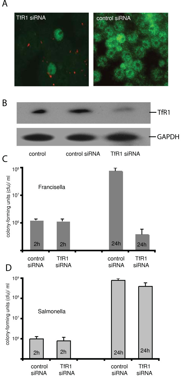
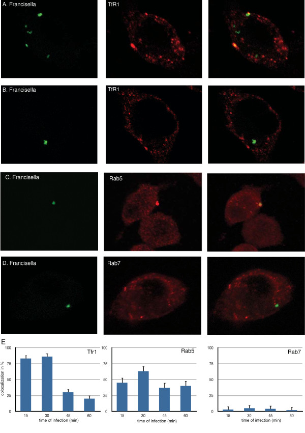
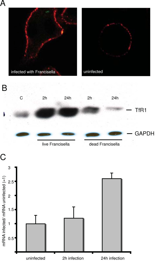
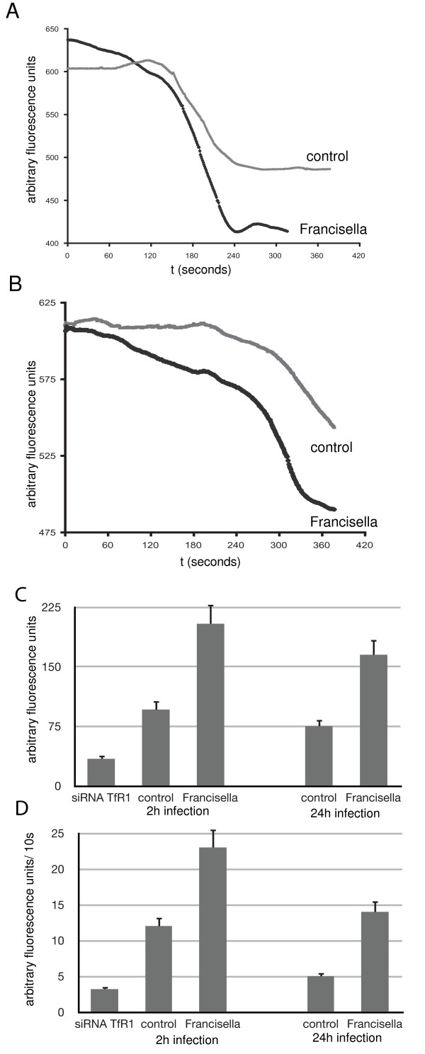
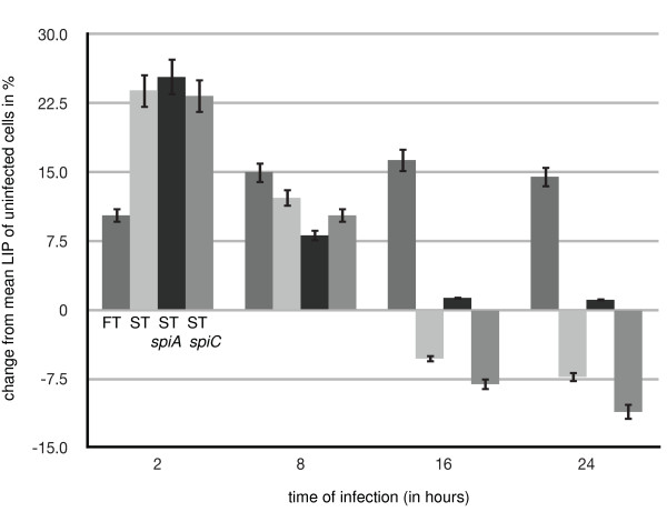
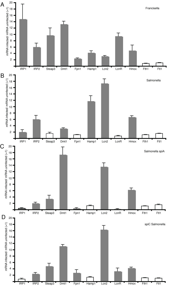
Results: We identified two distinct iron-handling scenarios for two different bacterial pathogens. Francisella tularensis drives an active iron acquisition program via the TfR1 pathway program with induction of ferrireductase (Steap3), iron membrane transporter Dmt1, and iron regulatory proteins IRP1 and IRP2, which is associated with a sustained increase of the labile iron pool inside the macrophage. Expression of TfR1 is critical for Francisella's intracellular proliferation. This contrasts with infection of macrophages by wild-type Salmonella typhimurium, which does not require expression of TfR1 for successful intracellular survival. Macrophages infected with Salmonella lack significant induction of Dmt1, Steap3, and IRP1, and maintain their labile iron pool at normal levels.

Conclusion: The distinction between two different phenotypes of iron utilization by intracellular pathogens will allow further characterization and understanding of host-cell iron metabolism and its modulation by intracellular bacteria.


Figures

Figure 1
Figure 1
Francisella, but not Salmonella requires TfR1 for proliferation inside macrophages. A. RAW264.7 macrophages were transfected with siRNA (coupled to Alexa Fluor 555, red fluorescence) specific for TfR1 or as control with random siRNA (no red fluorescence). After 48 h cells were fixed and processed for immunofluorescence with a mouse anti-TfR1 antibody followed by an Alexa488 conjugated goat-anti-mouse IgG (green fluorescence). Overlay of both fluorescence channels is shown. B. Proteins were solubilized from transfected and infected cells as above, separated on a 9% SDS-PAGE, transferred to Westran membranes, and immunoblotted with antiserum to TfR1. Visualization was by chemiluminescence C. RAW264.7 macrophages were transfected with TfR1-siRNA or with random siRNA (control). 48 h cells after transfection cells were infected with Francisella for 2 h or 24 h. The number of intracellular bacteria was obtained by plating a lysate of the host cells on chocolate agar plates for colony-forming units (cfus). Means of triplicate experiments +/- 1 standard error of mean are shown. D. RAW264.7 cells were treated as in C and then infected with Salmonella for 2 h or 24 h. The number of intracellular bacteria was determined as in C. Means of triplicate experiments +/- 1 standard error of mean are shown.
Figure 2
Figure 2
Transferrin receptor TfR1 and Rab5, but not Rab7, co-localize with Francisella. Macrophages (RAW264.7) were infected with Francisella that constitutively expressed green fluorescence protein (Gfp). At defined time intervals of infection, cells were fixed and stained with goat anti-TfR1 (A, B), with rabbit anti-Rab5 (C), or goat anti-Rab7 (D), followed by reaction with goat-anti-rabbit or rabbit-anti-goat IgG conjugated to Alexa594 (red fluorescence). Representative confocal images for thirty minutes of infection from twenty z-stacks acquired at 0.2 μm intervals are shown for each fluorescence channel, which were then merged using Volocity 4.1 software package (Improvision). E. The colocalization of Francisella with TfR1, Rab5, or Rab7 is described quantitatively for each time point by analyzing 100 infected cells from triplicate independent infection experiments. Means +/- 1 standard error of mean (SEM) are shown.
Figure 3
Figure 3
Infection with Francisella increases expression of transferrin receptor. A. RAW264.7 macrophages were infected with Francisella that constitutively expressed Gfp. After 2 h infected cells were fixed and processed for immunofluorescence with a mouse anti-TfR1 antibody followed by an Alexa594 conjugated goat-anti-mouse IgG (red fluorescence). Single confocal planes for merged fluorescence channels are shown. B. RAW264.7 cells were infected with live or formalin-inactivated Francisella (dead) for two and twenty-four hours. Immunoblotting of solubilized proteins was done with mouse anti-TfR1 and mouse anti-GAPDH as control. Visualization was by chemiluminescence. C. mRNA levels for TfR1 in RAW264.7 macrophages were determined after 2 or 24 h of infection with Francisella by quantitative light cycler PCR; levels are normalized to GAPDH-mRNA levels. Means of n = 6 experiments +/- 1 standard error of mean (SEM) are shown.
Figure 4
Figure 4
Transferrin-mediated delivery of iron increases the labile iron pool in Francisella-infected cells more efficiently than in uninfected cells. RAW macrophages were infected with Francisella LVS for 2 h (A) or 24 h (B) or left uninfected (control) and then loaded with Calcein-AM. The cell suspension was maintained at 37°C in a fluorometer. After stabilization of the fluorescence signal, holo-transferrin was added to the solution (t = 0) and the fluorescence signal recorded at one-second intervals. A decrease in the fluorescence indicates chelation of incoming iron with calcein, the amount of which is proportional to the slope and amplitude of the fluorescence signal. Results of triplicate measurements from triplicate experiments (n = 9) as described in A and B were analyzed for total amount of iron acquired as measured by arbitrary fluorescence units (C) and velocity of iron acquisition as measured by the change of fluorescence over time (D). Total iron and rate of iron uptake was also analyzed for macrophages whose TfR1 expression was suppressed by siRNA (siRNA TfR1 in Figure 4C and 4D). Measurements were made 24 h after transfection of uninfected macrophages (RAW264.7) with siRNA. All Values are given as means +/- 1 standard error of mean (SEM).
Figure 5
Figure 5
Labile iron pool in macrophages during infection with Francisella and Salmonella. RAW264.7 macrophages were infected for 2 h, 8 h, 16 h, and 24 h with wild Francisella (FT), wild-type Salmonella (ST), spiA Salmonella (ST/spiA), or spiC Salmonella (ST/spiC). Labile iron pool was determined with the calcein method as described in detail in Materials and Methods. Measurements were in arbitrary fluorescence units standardized to uninfected samples. Data shown are the deviation in percentage from uninfected samples from triplicate experiments. Results are expressed as means +/- 1 standard error of mean (SEM).
Figure 6
Figure 6
Expression of genes involved in iron homeostasis during infection with Francisella or Salmonella. RAW264.7 macrophages were infected for 24 h with wild-type Francisella (A), wild type Salmonella (B), spiC Salmonella (C), or spiA Salmonella (D). Quantitative mRNA levels were determined by quantitative light cycler PCR for: iron-regulatory protein 1 (IRP1), iron regulatory protein 2 (IRP2), ferrireductase (Steap3), transmembrane iron transporter (Dmt1), lipocalin (Lcn2), lipocalin receptor (LcnR), ferroportin (Fpn1), antimicrobial peptide hepcidin (Hamp1), heme oxygenase (Hmox1), ferritin heavy chain 1(Fth1), ferritin light chain 1 (Ftl1), and ferritin light chain 2 (Ftl2). Measurements were standardized to GAPDH-mRNA levels for each experiment. Values shown represent the ratio of mRNA for a given gene in infected cells divided by the mRNA level in uninfected cells (mRNA infected/mRNA uninfected). Statistically significant expression data are shown by solid bars (Student's t-test, p < 0.05 is considered as significant; individual p-values are given in the text). Results from n = 6 experiments are expressed as means +/- 1 standard error of mean (SEM).

Similar articles

Cited by

References

    1. Radtke AL, O'Riordan MX. Intracellular innate resistance to bacterial pathogens. Cell Microbiol. 2006;8:1720–1729. doi: 10.1111/j.1462-5822.2006.00795.x. - DOI - PubMed
    1. Paradkar P, De Domenico I, Durchfort N, Zohn I, Kaplan J, Ward DM. Iron-depletion limits intracellular bacterial growth in macrophages. Blood. 2008;112:866–874. doi: 10.1182/blood-2007-12-126854. - DOI - PMC - PubMed
    1. Collins HL. The role of iron in infections with intracellular bacteria. Immunol Lett. 2003;85:193–195. doi: 10.1016/S0165-2478(02)00229-8. - DOI - PubMed
    1. Chlosta S, Fishman DS, Harrington L, Johnson EE, Knutson MD, Wessling-Resnick M, Cherayil BJ. The iron efflux protein ferroportin regulates the intracellular growth of Salmonella enterica. Infect Immun. 2006;74:3065–3067. doi: 10.1128/IAI.74.5.3065-3067.2006. - DOI - PMC - PubMed
    1. Bullen JJ, Rogers HJ, Spalding PB, Ward CG. Natural resistance, iron and infection: a challenge for clinical medicine. J Med Microbiol. 2006;55:251–258. doi: 10.1099/jmm.0.46386-0. - DOI - PubMed

Publication types

MeSH terms