

JAM-A is a novel surface marker for NG2-Glia in the adult mouse brain
- PMID: 20184779
- PMCID: PMC2837050
- DOI: 10.1186/1471-2202-11-27
JAM-A is a novel surface marker for NG2-Glia in the adult mouse brain
Abstract
Background: Junctional adhesion molecule-A (JAM-A) is an adhesive protein expressed in various cell types. JAM-A localizes to the tight junctions between contacting endothelial and epithelial cells, where it contributes to cell-cell adhesion and to the control of paracellular permeability.
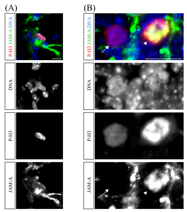
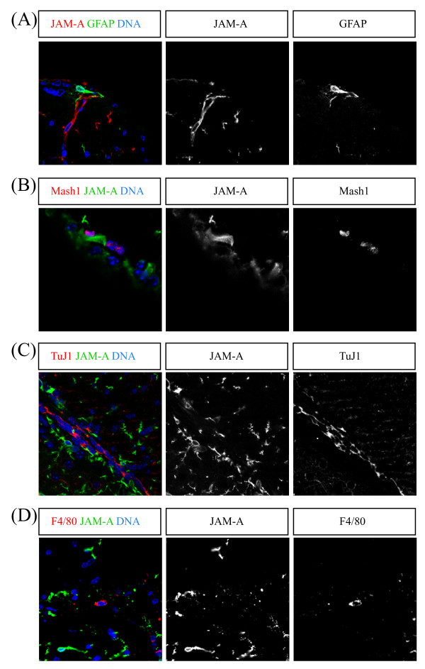
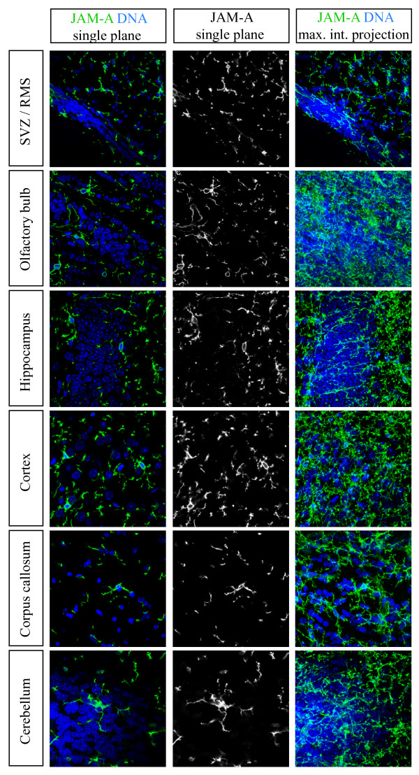
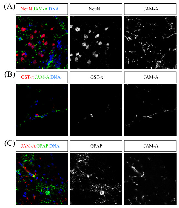
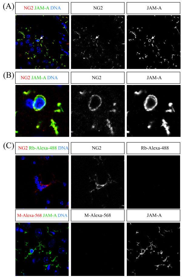
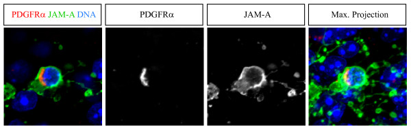
Results: So far, the expression pattern of JAM-A has not been described in detail for the different cell types of the adult brain. Here we show that a subset of proliferating cells in the adult mouse brain express JAM-A. We further clarify that these cells belong to the lineage of NG2-glia cells. Although these mitotic NG2-glia cells express JAM-A, the protein never shows a polarized subcellular distribution. Also non-mitotic NG2-glia cells express JAM-A in a non-polarized pattern on their surface.
Conclusions: Our data show that JAM-A is a novel surface marker for NG2-glia cells of the adult brain.
Figures
Similar articles
-
Astrocytes and NG2-glia: what's in a name?J Anat. 2005 Dec;207(6):687-93. doi: 10.1111/j.1469-7580.2005.00489.x. J Anat. 2005. PMID: 16367796 Free PMC article. Review.
-
AN2/NG2 protein-expressing glial progenitor cells in the murine CNS: isolation, differentiation, and association with radial glia.Glia. 2001 May;34(3):213-28. doi: 10.1002/glia.1055. Glia. 2001. PMID: 11329183
-
Morphological and physiological interactions of NG2-glia with astrocytes and neurons.J Anat. 2007 Jun;210(6):661-70. doi: 10.1111/j.1469-7580.2007.00729.x. Epub 2007 Apr 25. J Anat. 2007. PMID: 17459143 Free PMC article. Review.
-
NG2-expressing cells in the nervous system: role of the proteoglycan in migration and glial-neuron interaction.J Anat. 2005 Dec;207(6):735-44. doi: 10.1111/j.1469-7580.2005.00461.x. J Anat. 2005. PMID: 16367801 Free PMC article. Review.
-
Mixed primary culture and clonal analysis provide evidence that NG2 proteoglycan-expressing cells after spinal cord injury are glial progenitors.Dev Neurobiol. 2007 Jun;67(7):860-74. doi: 10.1002/dneu.20369. Dev Neurobiol. 2007. PMID: 17506499
Cited by
-
High-throughput flow cytometry screening reveals a role for junctional adhesion molecule a as a cancer stem cell maintenance factor.Cell Rep. 2014 Jan 16;6(1):117-29. doi: 10.1016/j.celrep.2013.11.043. Epub 2013 Dec 27. Cell Rep. 2014. PMID: 24373972 Free PMC article.
-
The F11 Receptor (F11R)/Junctional Adhesion Molecule-A (JAM-A) (F11R/JAM-A) in cancer progression.Mol Cell Biochem. 2022 Jan;477(1):79-98. doi: 10.1007/s11010-021-04259-2. Epub 2021 Sep 17. Mol Cell Biochem. 2022. PMID: 34533648 Free PMC article. Review.
-
Expression and prognostic value of JAM-A in gliomas.J Neurooncol. 2017 Oct;135(1):107-117. doi: 10.1007/s11060-017-2555-0. Epub 2017 Jul 4. J Neurooncol. 2017. PMID: 28677106 Free PMC article.
-
The NG2 Proteoglycan Protects Oligodendrocyte Precursor Cells against Oxidative Stress via Interaction with OMI/HtrA2.PLoS One. 2015 Sep 4;10(9):e0137311. doi: 10.1371/journal.pone.0137311. eCollection 2015. PLoS One. 2015. PMID: 26340347 Free PMC article.
References
Publication types
MeSH terms
Substances
LinkOut - more resources
Full Text Sources
