

Neural circuitry of emotional face processing in autism spectrum disorders
- PMID: 20184808
- PMCID: PMC2834792
- DOI: 10.1503/jpn.090085
Neural circuitry of emotional face processing in autism spectrum disorders
Abstract
Background: Autism spectrum disorders (ASD) are associated with severe impairments in social functioning. Because faces provide nonverbal cues that support social interactions, many studies of ASD have examined neural structures that process faces, including the amygdala, ventromedial prefrontal cortex and superior and middle temporal gyri. However, increases or decreases in activation are often contingent on the cognitive task. Specifically, the cognitive domain of attention influences group differences in brain activation. We investigated brain function abnormalities in participants with ASD using a task that monitored attention bias to emotional faces.
Methods: Twenty-four participants (12 with ASD, 12 controls) completed a functional magnetic resonance imaging study while performing an attention cuing task with emotional (happy, sad, angry) and neutral faces.
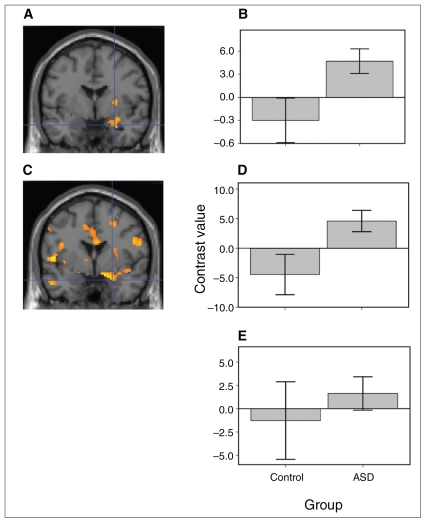
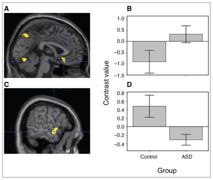
Results: In response to emotional faces, those in the ASD group showed greater right amygdala activation than those in the control group. A preliminary psychophysiological connectivity analysis showed that ASD participants had stronger positive right amygdala and ventromedial prefrontal cortex coupling and weaker positive right amygdala and temporal lobe coupling than controls. There were no group differences in the behavioural measure of attention bias to the emotional faces.
Limitations: The small sample size may have affected our ability to detect additional group differences.
Conclusion: When attention bias to emotional faces was equivalent between ASD and control groups, ASD was associated with greater amygdala activation. Preliminary analyses showed that ASD participants had stronger connectivity between the amygdala ventromedial prefrontal cortex (a network implicated in emotional modulation) and weaker connectivity between the amygdala and temporal lobe (a pathway involved in the identification of facial expressions, although areas of group differences were generally in a more anterior region of the temporal lobe than what is typically reported for emotional face processing). These alterations in connectivity are consistent with emotion and face processing disturbances in ASD.
Figures

Similar articles
-
Amygdala habituation and prefrontal functional connectivity in youth with autism spectrum disorders.J Am Acad Child Adolesc Psychiatry. 2013 Jan;52(1):84-93. doi: 10.1016/j.jaac.2012.10.012. Epub 2012 Dec 2. J Am Acad Child Adolesc Psychiatry. 2013. PMID: 23265636 Free PMC article.
-
Neural activation to emotional faces in adolescents with autism spectrum disorders.J Child Psychol Psychiatry. 2011 Mar;52(3):296-305. doi: 10.1111/j.1469-7610.2010.02317.x. Epub 2010 Oct 7. J Child Psychol Psychiatry. 2011. PMID: 21039484 Free PMC article.
-
Early neural activation during facial affect processing in adolescents with Autism Spectrum Disorder.Neuroimage Clin. 2014 Nov 18;7:203-12. doi: 10.1016/j.nicl.2014.11.009. eCollection 2015. Neuroimage Clin. 2014. PMID: 25610782 Free PMC article.
-
Research review: Constraining heterogeneity: the social brain and its development in autism spectrum disorder.J Child Psychol Psychiatry. 2011 Jun;52(6):631-44. doi: 10.1111/j.1469-7610.2010.02349.x. Epub 2011 Jan 19. J Child Psychol Psychiatry. 2011. PMID: 21244421 Free PMC article. Review.
-
Functional atlas of emotional faces processing: a voxel-based meta-analysis of 105 functional magnetic resonance imaging studies.J Psychiatry Neurosci. 2009 Nov;34(6):418-32. J Psychiatry Neurosci. 2009. PMID: 19949718 Free PMC article. Review.
Cited by
-
Support vector machine prediction of individual Autism Diagnostic Observation Schedule (ADOS) scores based on neural responses during live eye-to-eye contact.Sci Rep. 2024 Feb 8;14(1):3232. doi: 10.1038/s41598-024-53942-z. Sci Rep. 2024. PMID: 38332184 Free PMC article.
-
Age-related abnormalities in white matter microstructure in autism spectrum disorders.Brain Res. 2012 Oct 15;1479:1-16. doi: 10.1016/j.brainres.2012.07.056. Epub 2012 Aug 10. Brain Res. 2012. PMID: 22902768 Free PMC article.
-
Amygdala habituation and prefrontal functional connectivity in youth with autism spectrum disorders.J Am Acad Child Adolesc Psychiatry. 2013 Jan;52(1):84-93. doi: 10.1016/j.jaac.2012.10.012. Epub 2012 Dec 2. J Am Acad Child Adolesc Psychiatry. 2013. PMID: 23265636 Free PMC article.
-
Increased Functional Connectivity During Emotional Face Processing in Children With Autism Spectrum Disorder.Front Hum Neurosci. 2018 Oct 10;12:408. doi: 10.3389/fnhum.2018.00408. eCollection 2018. Front Hum Neurosci. 2018. PMID: 30364114 Free PMC article.
-
Differences in cortical processing of facial emotions in broader autism phenotype.PLoS One. 2022 Jan 18;17(1):e0262004. doi: 10.1371/journal.pone.0262004. eCollection 2022. PLoS One. 2022. PMID: 35041646 Free PMC article. Clinical Trial.
References
-
- American Psychiatric Association. Diagnostic and statistical manual of mental disorders. 4th ed. Washington (DC): the Association; 1994.
-
- Adolphs R. The neurobiology of social cognition. Curr Opin Neurobiol. 2001;11:231–9. - PubMed
-
- Baron-Cohen S, Ring HA, Bullmore ET, et al. The amygdala theory of autism. Neurosci Biobehav Rev. 2000;24:355–64. - PubMed
-
- Schultz RT. Developmental deficits in social perception in autism: the role of the amygdala and fusiform face area. Int J Dev Neurosci. 2005;23:125–41. - PubMed
-
- Ashwin C, Baron-Cohen S, Wheelwright S, et al. Differential activation of the amygdala and the “social brain” during fearful face-processing in Asperger Syndrome. Neuropsychologia. 2007;45:2–14. - PubMed
Publication types
MeSH terms
Grants and funding
LinkOut - more resources
Full Text Sources
Other Literature Sources