

Longitudinal study of carbon monoxide intoxication by diffusion tensor imaging with neuropsychiatric correlation
- PMID: 20184809
- PMCID: PMC2834793
- DOI: 10.1503/jpn.090057
Longitudinal study of carbon monoxide intoxication by diffusion tensor imaging with neuropsychiatric correlation
Abstract
Background: White matter damage is common after carbon monoxide (CO) intoxication, but in vivo follow-up studies about the mechanism of white matter damage are not possible in pathology series. Diffusion tensor imaging (DTI) and voxel-based morphometry (VBM) can quantify diffusion parameters and volumetric changes in white matter that can be correlated with neuropsychological performances in longitudinal studies.
Methods: We examined 9 patients with CO intoxication using DTI, VBM and neuropsychologic tests at an average of 3 and 10 months after CO exposure. We used data from 18 age- and sex-matched controls for comparison.
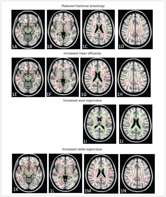
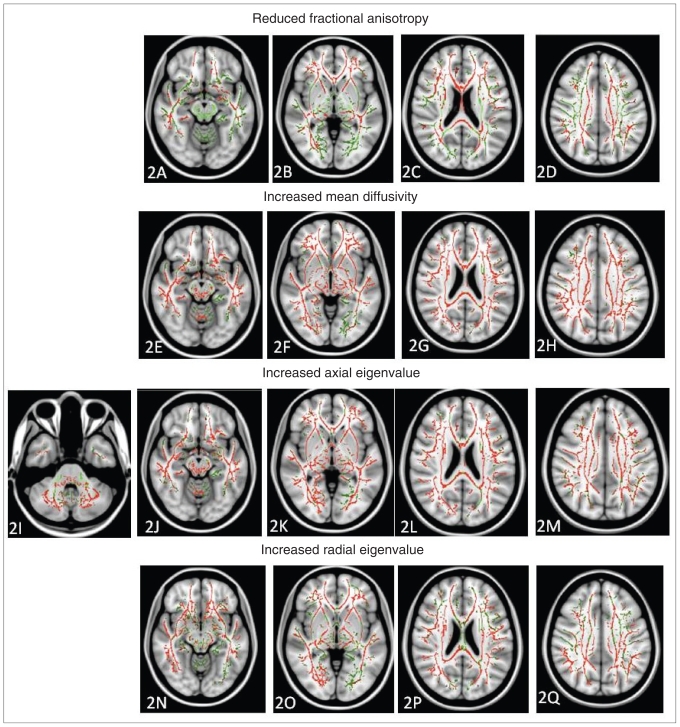
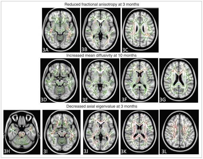
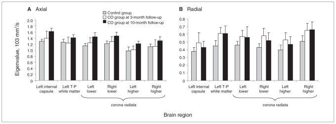
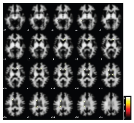
Results: We found that cognitive recovery at 10 months after CO intoxication was not significant, although it was after 3 months. The neuropsychologic tests correlated better for the fibre tract of the semicentrum ovale and not the periventricular fibres. Diffusion measures suggest increases in fractional anisotropy, mean diffusivity and axial eigenvalues over time, while increases in radial eigenvalue were evident at 3 months compared with controls. Periventricular white matter atrophy was observed 10 months after CO intoxication.
Limitations: Our study included few cases, and the interpretation of the putative changes on neuroimaging findings cannot be confirmed by histology.
Conclusion: Our study showed that the evolution of white matter injury in CO encephalopathy occurred over time. Cognitive recovery was not evident in the follow-up period because of white matter injuries.
Figures
Similar articles
-
Damage of white matter tract correlated with neuropsychological deficits in carbon monoxide intoxication after hyperbaric oxygen therapy.J Neurotrauma. 2009 Aug;26(8):1263-70. doi: 10.1089/neu.2008.0619. J Neurotrauma. 2009. PMID: 19317622
-
Assessment of damage to cerebral white matter fiber in the subacute phase after carbon monoxide poisoning using fractional anisotropy in diffusion tensor imaging.Neuroradiology. 2010 Aug;52(8):735-43. doi: 10.1007/s00234-009-0649-x. Epub 2010 Jan 12. Neuroradiology. 2010. PMID: 20066405
-
Callosal damage and cognitive deficits in chronic carbon monoxide intoxication: A diffusion tensor imaging study.J Neurol Sci. 2015 Aug 15;355(1-2):101-7. doi: 10.1016/j.jns.2015.05.030. Epub 2015 May 28. J Neurol Sci. 2015. PMID: 26033717
-
White matter hyperintensities and neuropsychological outcome following carbon monoxide poisoning.Neurology. 2002 May 28;58(10):1525-32. doi: 10.1212/wnl.58.10.1525. Neurology. 2002. PMID: 12034791 Review.
-
Cerebral white matter integrity and cognitive aging: contributions from diffusion tensor imaging.Neuropsychol Rev. 2009 Dec;19(4):415-35. doi: 10.1007/s11065-009-9113-2. Epub 2009 Aug 25. Neuropsychol Rev. 2009. PMID: 19705281 Free PMC article. Review.
Cited by
-
The role of MR imaging in assessment of brain damage from carbon monoxide poisoning: a review of the literature.AJNR Am J Neuroradiol. 2014 Apr;35(4):625-31. doi: 10.3174/ajnr.A3489. Epub 2013 Apr 18. AJNR Am J Neuroradiol. 2014. PMID: 23598831 Free PMC article. Review.
-
Longitudinal White Matter Changes following Carbon Monoxide Poisoning: A 9-Month Follow-Up Voxelwise Diffusional Kurtosis Imaging Study.AJNR Am J Neuroradiol. 2019 Mar;40(3):478-482. doi: 10.3174/ajnr.A5979. Epub 2019 Feb 14. AJNR Am J Neuroradiol. 2019. PMID: 30765380 Free PMC article.
-
Hyperbaric Oxygen Therapy for Carbon Monoxide-Induced Delayed Neuropsychiatric Sequelae: Case Report of Two Cases and Relevant Literature Review.Case Rep Psychiatry. 2021 Mar 2;2021:6663824. doi: 10.1155/2021/6663824. eCollection 2021. Case Rep Psychiatry. 2021. PMID: 33763276 Free PMC article.
-
Heterogeneous Diffusion Metrics Patterns in Delayed Encephalopathy After Acute Carbon Monoxide Poisoning: A Case Report.Brain Neurorehabil. 2023 Nov 6;16(3):e34. doi: 10.12786/bn.2023.16.e34. eCollection 2023 Nov. Brain Neurorehabil. 2023. PMID: 38047103 Free PMC article.
-
Impact of Hyperbaric Oxygen Therapy on Cognitive Functions: a Systematic Review.Neuropsychol Rev. 2022 Mar;32(1):99-126. doi: 10.1007/s11065-021-09500-9. Epub 2021 Apr 13. Neuropsychol Rev. 2022. PMID: 33847854 Free PMC article.
References
-
- Kuo CJ, Conwell Y, Yu Q, et al. Suicide by charcoal burning in Taiwan: implications for means substitution by a case-linkage study. Soc Psychiatry Psychiatr Epidemiol. 2008;43:286–90. - PubMed
-
- Young LJ, Caughey WS. Pathobiochemistry of CO poisoning. FEBS Lett. 1990;272:1–6. - PubMed
-
- Weaver LK. Clinical practice. Carbon monoxide poisoning. N Engl J Med. 2009;360:1217–25. - PubMed
-
- Hopkins RO, Fearing MA, Weaver LK, et al. Basal ganglia lesions following carbon monoxide poisoning. Brain Inj. 2006;20:273–81. - PubMed
Publication types
MeSH terms
LinkOut - more resources
Full Text Sources
Medical