Mutant huntingtin fragment selectively suppresses Brn-2 POU domain transcription factor to mediate hypothalamic cell dysfunction
- PMID: 20185558
- PMCID: PMC2865370
- DOI: 10.1093/hmg/ddq087
Mutant huntingtin fragment selectively suppresses Brn-2 POU domain transcription factor to mediate hypothalamic cell dysfunction
Abstract
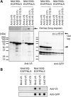
In polyglutamine diseases including Huntington's disease (HD), mutant proteins containing expanded polyglutamine stretches form nuclear aggregates in neurons. Although analysis of their disease models suggested a significance of transcriptional dysregulation in these diseases, how it mediates the specific neuronal cell dysfunction remains obscure. Here we performed a comprehensive analysis of altered DNA binding of multiple transcription factors using R6/2 HD model mice brains that express an N-terminal fragment of mutant huntingtin (mutant Nhtt). We found a reduction of DNA binding of Brn-2, a POU domain transcription factor involved in differentiation and function of hypothalamic neurosecretory neurons. We provide evidence supporting that Brn-2 loses its function through two pathways, its sequestration by mutant Nhtt and its reduced transcription, leading to reduced expression of hypothalamic neuropeptides. In contrast to Brn-2, its functionally related protein, Brn-1, was not sequestered by mutant Nhtt but was upregulated in R6/2 brain, except in hypothalamus. Our data indicate that functional suppression of Brn-2 together with a region-specific lack of compensation by Brn-1 mediates hypothalamic cell dysfunction by mutant Nhtt.
Figures


Similar articles
-
Selection of behaviors and segmental coordination during larval locomotion is disrupted by nuclear polyglutamine inclusions in a new Drosophila Huntington's disease-like model.J Neurogenet. 2010 Dec;24(4):194-206. doi: 10.3109/01677063.2010.514367. J Neurogenet. 2010. PMID: 21087194
-
Mutant Huntingtin reduces HSP70 expression through the sequestration of NF-Y transcription factor.EMBO J. 2008 Mar 19;27(6):827-39. doi: 10.1038/emboj.2008.23. Epub 2008 Feb 21. EMBO J. 2008. PMID: 18288205 Free PMC article.
-
Lack of huntingtin-associated protein-1 causes neuronal death resembling hypothalamic degeneration in Huntington's disease.J Neurosci. 2003 Jul 30;23(17):6956-64. doi: 10.1523/JNEUROSCI.23-17-06956.2003. J Neurosci. 2003. PMID: 12890790 Free PMC article.
-
Nature and cause of mitochondrial dysfunction in Huntington's disease: focusing on huntingtin and the striatum.J Neurochem. 2010 Jul;114(1):1-12. doi: 10.1111/j.1471-4159.2010.06741.x. Epub 2010 Apr 9. J Neurochem. 2010. PMID: 20403078 Review.
-
Mechanisms for neuronal cell death and dysfunction in Huntington's disease: pathological cross-talk between the nucleus and the mitochondria?J Mol Med (Berl). 2001 Jul;79(7):375-81. doi: 10.1007/s001090100223. J Mol Med (Berl). 2001. PMID: 11466559 Review.
Cited by
-
Could yeast prion domains originate from polyQ/N tracts?Prion. 2013 May-Jun;7(3):209-14. doi: 10.4161/pri.24628. Prion. 2013. PMID: 23764835 Free PMC article.
-
POU-III transcription factors (Brn1, Brn2, and Oct6) influence neurogenesis, molecular identity, and migratory destination of upper-layer cells of the cerebral cortex.Cereb Cortex. 2013 Nov;23(11):2632-43. doi: 10.1093/cercor/bhs252. Epub 2012 Aug 14. Cereb Cortex. 2013. PMID: 22892427 Free PMC article.
-
Oxytocin Prevents the Development of 3-NP-Induced Anxiety and Depression in Male and Female Rats: Possible Interaction of OXTR and mGluR2.Cell Mol Neurobiol. 2022 May;42(4):1105-1123. doi: 10.1007/s10571-020-01003-0. Epub 2020 Nov 17. Cell Mol Neurobiol. 2022. PMID: 33201416 Free PMC article.
-
Thalamocortical axons regulate neurogenesis and laminar fates in the early sensory cortex.Proc Natl Acad Sci U S A. 2022 May 31;119(22):e2201355119. doi: 10.1073/pnas.2201355119. Epub 2022 May 25. Proc Natl Acad Sci U S A. 2022. PMID: 35613048 Free PMC article.
-
Hypothalamic Alterations in Neurodegenerative Diseases and Their Relation to Abnormal Energy Metabolism.Front Mol Neurosci. 2018 Jan 19;11:2. doi: 10.3389/fnmol.2018.00002. eCollection 2018. Front Mol Neurosci. 2018. PMID: 29403354 Free PMC article. Review.
References
-
- Walker F.O. Huntington's disease. Lancet. 2007;369:218–228. doi:10.1016/S0140-6736(07)60111-1. - DOI - PubMed
-
- Landles C., Bates G.P. Huntingtin and the molecular pathogenesis of Huntington's disease. Fourth in molecular medicine review series. EMBO Rep. 2004;5:958–963. doi:10.1038/sj.embor.7400250. - DOI - PMC - PubMed
-
- Bauer P.O., Nukina N. The pathogenic mechanisms of polyglutamine diseases and current therapeutic strategies. J. Neurochem. 2009;110:1737–1765. doi:10.1111/j.1471-4159.2009.06302.x. - DOI - PubMed
-
- Luthi-Carter R., Hanson S.A., Strand A.D., Bergstrom D.A., Chun W., Peters N.L., Woods A.M., Chan E.Y., Kooperberg C., Krainc D., et al. Dysregulation of gene expression in the R6/2 model of polyglutamine disease: parallel changes in muscle and brain. Hum. Mol. Genet. 2002;11:1911–1926. doi:10.1093/hmg/11.17.1911. - DOI - PubMed
-
- Chan E.Y., Luthi-Carter R., Strand A., Solano S.M., Hanson S.A., DeJohn M.M., Kooperberg C., Chase K.O., DiFiglia M., Young A.B., et al. Increased huntingtin protein length reduces the number of polyglutamine-induced gene expression changes in mouse models of Huntington's disease. Hum. Mol. Genet. 2002;11:1939–1951. doi:10.1093/hmg/11.17.1939. - DOI - PubMed
Publication types
MeSH terms
Substances
LinkOut - more resources
Full Text Sources
Medical
Molecular Biology Databases

