. 2010 Jun;38(11):3643-54.
doi: 10.1093/nar/gkq078. Epub 2010 Feb 25.

DNA linking number change induced by sequence-specific DNA-binding proteins

Affiliations

DNA linking number change induced by sequence-specific DNA-binding proteins

Bo Chen et al. Nucleic Acids Res. 2010 Jun.

Abstract

Sequence-specific DNA-binding proteins play a key role in many fundamental biological processes, such as transcription, DNA replication and recombination. Very often, these DNA-binding proteins introduce structural changes to the target DNA-binding sites including DNA bending, twisting or untwisting and wrapping, which in many cases induce a linking number change (Delta Lk) to the DNA-binding site. Due to the lack of a feasible approach, Delta Lk induced by sequence-specific DNA-binding proteins has not been fully explored. In this paper we successfully constructed a series of DNA plasmids that carry many tandem copies of a DNA-binding site for one sequence-specific DNA-binding protein, such as lambda O, LacI, GalR, CRP and AraC. In this case, the protein-induced Delta Lk was greatly amplified and can be measured experimentally. Indeed, not only were we able to simultaneously determine the protein-induced Delta Lk and the DNA-binding constant for lambda O and GalR, but also we demonstrated that the protein-induced Delta Lk is an intrinsic property for these sequence-specific DNA-binding proteins. Our results also showed that protein-mediated DNA looping by AraC and LacI can induce a Delta Lk to the plasmid DNA templates. Furthermore, we demonstrated that the protein-induced Delta Lk does not correlate with the protein-induced DNA bending by the DNA-binding proteins.


Figures

Figure 1.
Figure 1.
Plasmids used to determine protein-induced ΔLk by λ O protein, GalR, CRP, AraC and LacI. Plasmids pCB34, pCB46 and pCB55 were derived from pACYC184; plasmids pYZX36, pYZX42 and pYZX46 were derived from pACYCDuet-1. These plasmids were constructed as detailed under ‘Materials and Methods’ section. The restriction enzyme sites for BamHI, BglII, XhoI, AvrII, Nb.BsrDI and Nt.BbvCI are shown. The small closed rectangles represent the site specific recognition sequence for λ O protein (Iteron III), GalR (gal OE), CRP, AraC (araI) and LacI (lacO1).
Figure 2.
Figure 2.
Simultaneous determination of λ O protein-induced DNA unwinding angle (β) or ΔLk and the DNA-binding constant (K). (A) and (C) λ O protein titration assays were performed as described under ‘Materials and Methods’ section using the ligation method (A) or the Topo I method (C). In addition to 0.0625 nM of plasmid pCB5, the reaction mixtures for the DNA samples applied to lanes 1–10 also contain 0, 1.5, 2.0, 3.0, 4.0, 5.0, 6.3, 7.5, 10.0, 12.5 nM of λ O protein (as dimer), respectively. DNA topoisomers were resolved with electrophoresis in 1% agarose gel containing 0.5 µg/ml chloroquine. (B) and (D) Quantification analysis of the binding data from the λ O titration experiments. β and K were calculated according to Equation 1. ΔLk was calculated by Equation 2.
Figure 3.
Figure 3.
The Determination of the DNA-binding constant (K) and the protein-induced DNA bending angle for λ O protein. The EMSA assays and the DNA bending assays were performed as described under ‘Materials and Methods’ section. (A) Binding of λ O protein to the DNA oligomer containing the iteron III of coliphage λ DNA replication origin. A 145 bp 32P-labeled EcoRV fragment of pBend2-Iteron III was incubated with increasing concentrations of λ O protein in 50 µl of 1× EMSA binding buffer containing 10 mM Tris–HCl (pH 7.4), 20 mM KCl, 0.5 mM EDTA, 1 mM DTT, 5 mM MgCl2 and 5% glycerol. The autoradiogram was shown. The radioactivities were quantified with a PhosphorImager. Lane 1 is free DNA fragment. In addition to the 32P-labeled DNA fragment, the reaction mixtures for the DNA samples applied to lanes 2–11 also contains 0, 1, 2.5, 5, 7.5, 10, 15, 20, 25, 50 and 100 nM of λ O protein (as dimer), respectively. (B) Quantification analysis of the binding data from the EMSA experiments. The bound ratio of DNA was plotted against the protein concentration. The curve was generated by fitting the data to Equation 6 as described under ‘Materials and Methods’ section to yield the DNA-binding constant of 2.5 × 10⁸ M−1. (C) The DNA bending assay. After the binding of λ O protein to the permutated DNA fragments, an 8% polyacrylamide gel was used to separate the bound and free DNA fragments. The autoradiogram of 32P-labeled DNA fragments was shown. Lanes 1–5 are the λ iteron III fragments produced by digestion of plasmid pBend2-Iteron III with restriction enzymes of MluI, NheI, EcoRV, SspI and BamHI, respectively. The DNA fragments in the bottom of the gels are free DNA, and those in the upper part are protein–DNA complexes.
Figure 4.
Figure 4.
Simultaneous determination of GalR-induced DNA-unwinding angle (β) or ΔLk and the DNA-binding constant (K). GalR titration assays were performed as described in ‘Materials and Methods’ section using the ligation method. DNA topoisomers were resolved with electrophoresis in 1% agarose gel containing 0.5 µg/ml chloroquine. (A) The GalR titration experiment. In addition to 0.055 nM of plasmid pCB42, the reaction mixtures for the DNA samples applied to lanes 1–10 also contain 0, 1.3, 1.5, 2.0, 2.5, 3.0, 4.0, 6.0, 8.0, 10.0 nM of GalR (as dimer), respectively. (B) GalR failed to unwind the DNA when d-galactose was present. The reaction mixture for the DNA sample applied to lane 1 contains neither GalR nor d-galactose. As indicated at the top of the image, the reaction mixtures for the DNA samples applied to lanes 2–4 contain GalR (lanes 2 and 4, 5 nM; lanes 3 and 5, 10 nM) and D-galactose (lanes 4 and 5, 1 mM). (C) Quantification analysis of the binding data from GalR titration experiments. β and K were determined according to Equation 1. ΔLk was calculated by Equation 2.
Figure 5.
Figure 5.
AraC-induced ΔLk. Experiments to determine AraC-induced ΔLk were performed as described under ‘Materials and Methods’ section. DNA topoisomers were resolved with electrophoresis in 1% agarose gel containing 0.5 µg/ml chloroquine. (A) The AraC titration experiment. In addition to 0.0714 nM of plasmid pYZX42, the reaction mixtures for the DNA samples applied to lanes 1–11 also contain 0, 2.5, 5.0, 7.5, 10.0, 12.5, 15.0, 20.0, 25.0, 30.0, 35.0 nM of AraC (as dimer), respectively. (B) AraC failed to induce ΔLk of pYZX42 when l-arabinose was present. The reaction mixture for the DNA sample applied to lane 1 contains neither AraC nor l-arabinose. As indicated at the top of the image, the reaction mixtures for the DNA samples applied to lanes 2–5 contain AraC (lanes 2 and 4, 3 nM; lanes 3 and 5, 6 nM) and l-arabinose (lanes 4 and 5, 1 mM).
Figure 6.
Figure 6.
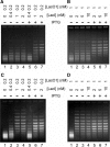
The LacI-induced ΔLk in the presence or absence of IPTG. The experiments to determine LacI-induced ΔLk were performed according to the ligation method (A and B) or the Topo I method (C and D) as described under ‘Materials and Methods’ section. Plasmid pYZX46 that contains 19 lac O1 operators was used as the DNA template. The concentration of lac O1 operator and LacI (as tetramer) are shown at the top of the images. The reaction mixtures for the DNA samples applied to lanes 5–7 also contain 1 mM of IPTG.
Figure 7.
Figure 7.
CRP-induced ΔLk, the DNA-bending angle (α), and the DNA-binding constant. (A) CRP titration experiments were performed according to the ligation method as described under ‘Materials and Methods’ section. In addition to 0.0417 nM of plasmid pCB51 and 20 µM of cAMP, the reaction mixtures for the DNA samples applied to lanes 1–10 also contain 0, 1.3, 1.5, 2.0, 2.5, 3.0, 4.0, 6.0, 8.0, 10.0 nM of CRP (as dimer), respectively. DNA topoisomers were resolved with electrophoresis in 1% agarose gel containing 0.5 µg/ml chloroquine. (B) DNA bending assays were carried out as described under ‘Materials and Methods’ section. After the binding of CRP to the permutated DNA fragments in the presence of 20 µM of cAMP, an 8% polyacrylamide gel was used to separate the bound and free DNA fragments. The autoradiogram of 32P-labeled DNA fragments was shown. Lanes 1–9 are the lac P1 promoter’s CRP fragments produced by digestion of plasmid pBend2-CRP with restriction enzymes of MluI, BglII, NheI, SpeI, XhoI, DraI, EcoRV, NruI and BamHI, respectively. The DNA fragments in the bottom of the gels are free DNA, and those in the upper part are protein-DNA complexes. (C) The EMSA assays were performed as described under ‘Materials and Methods’ section. A 159-bp 32P-labeled EcoRV fragment of pBEND2-CRP was incubated with increasing concentrations of CRP in 50 µl of 1 × EMSA binding buffer containing 20 mM Tris–HCl (pH 8.0), 20 µM cAMP, 200 mM NaCl, 0.5 mM EDTA, 1 mM DTT, 5 mM MgCl2 and 5% glycerol. The autoradiogram was shown. The radioactivities were quantified with a PhosphorImager. Lane 1 is free DNA fragment. In addition to the 32P-labeled DNA fragment, the reaction mixtures for the DNA samples applied to lanes 2–11 also contains 0.1, 0.25, 0.5, 1, 1.5, 2.0, 3.0, 5.0, 10.0 and 20.0 nM of CRP (as dimer), respectively. (D) Quantification analysis of the binding data from the EMSA experiments. The bound ratio of DNA was plotted against the protein concentration. The curve was generated by fitting the data to Equation 6 as described under ‘Materials and Methods’ section to yield the DNA-binding constant of 2.1 × 10⁹ M−1.
Figure 8.
Figure 8.
Proposed models to explain the protein-induced ΔLk by sequence-specific DNA-binding proteins. Models (A to B) show that certain site-specific DNA-wrapping proteins, such as λ O protein, bind and wrap the DNA recognition sites to induce the ΔLk of the DNA template. Models (C to D) demonstrate that DNA-looping proteins, such as AraC and LacI, form a topological nucleoprotein complex and therefore induce the ΔLk of the DNA template. Blue circle and red cylinder, respectively, represent the DNA recognition sequence for a site-specific DNA-binding protein and the site-specific DNA protein.

Similar articles

Cited by

References

    1. Kaguni JM. DnaA: controlling the initiation of bacterial DNA replication and more. Annu. Rev. Microbiol. 2006;60:351–375. - PubMed
    1. Zahn K, Blattner FR. Binding and bending of the lambda replication origin by the phage O protein. EMBO J. 1985;4:3605–3616. - PMC - PubMed
    1. Mott ML, Berger JM. DNA replication initiation: mechanisms and regulation in bacteria. Nat. Rev. Microbiol. 2007;5:343–354. - PubMed
    1. Leonard AC, Grimwade JE. Building a bacterial orisome: emergence of new regulatory features for replication origin unwinding. Mol. Microbiol. 2005;55:978–985. - PMC - PubMed
    1. Alfano C, McMacken R. Ordered assembly of nucleoprotein structures at the bacteriophage lambda replication origin during the initiation of DNA replication. J. Biol. Chem. 1989;264:10699–10708. - PubMed

Publication types

MeSH terms