. 2010 Apr;176(4):1927-40.
doi: 10.2353/ajpath.2010.090561. Epub 2010 Feb 25.

Ricinus communis agglutinin I leads to rapid down-regulation of VEGFR-2 and endothelial cell apoptosis in tumor blood vessels

Affiliations

Ricinus communis agglutinin I leads to rapid down-regulation of VEGFR-2 and endothelial cell apoptosis in tumor blood vessels

Weon-Kyoo You et al. Am J Pathol. 2010 Apr.

Abstract

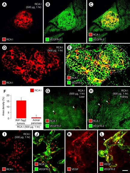
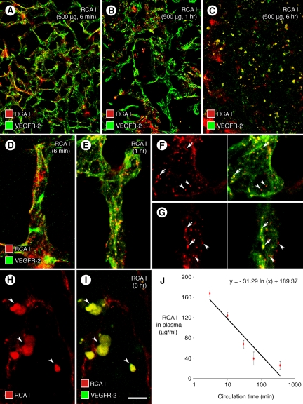
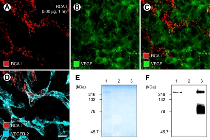
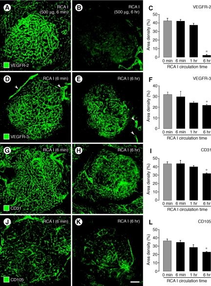
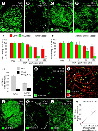
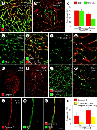
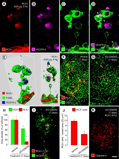
Ricinus communis agglutinin I (RCA I), a galactose-binding lectin from castor beans, binds to endothelial cells at sites of plasma leakage, but little is known about the amount and functional consequences of binding to tumor endothelial cells. We addressed this issue by examining the effects of RCA I on blood vessels of spontaneous pancreatic islet-cell tumors in RIP-Tag2 transgenic mice. After intravenous injection, RCA I bound strongly to tumor vessels but not to normal blood vessels. At 6 minutes, RCA I fluorescence of tumor vessels was largely diffuse, but over the next hour, brightly fluorescent dots appeared as the lectin was internalized by endothelial cells. RCA I injection led to a dose- and time-dependent decrease in vascular endothelial growth factor receptor-2 (VEGFR-2) immunoreactivity in tumor endothelial cells, with 95% loss over 6 hours. By comparison, VEGFR-3, CD31, and CD105 had decreases in the range of 21% to 33%. Loss of VEGFR-2 was followed by increased activated caspase-3 in tumor vessels. Prior inhibition of VEGF signaling by AG-028262 decreased RCA I binding and internalization into tumor vessels. These findings indicate RCA I preferentially binds to and is internalized by tumor endothelial cells, which leads to VEGFR-2 down-regulation, endothelial cell apoptosis, and tumor vessel regression. Together, the results illustrate the selective impact of RCA I on VEGF signaling in tumor blood vessels.


Figures

Figure 1
Figure 1
Preferential binding and internalization of RCA I by tumor vessels. Micrographs of RIP-Tag2 tumors (AC) showing strong red fluorescence of tumor vessels 1 hour after i.v. injection of 500 μg rhodamine-RCA I and green VEGFR-2 immunofluorescence. Strong red fluorescence of tumor vessels (D, E, dashed lines outline tumors) contrasts with little or no fluorescence of blood vessels in surrounding acinar pancreas. Bar graph (F) compares abundant RCA I fluorescence (fractional area, 15%) of tumor vessels with almost no fluorescence (0.6%) in the acinar pancreas. Micrographs show weak RCA I fluorescence in central veins of the liver (G, arrowheads) and glomeruli of the kidney (H, arrowheads). Confocal micrographs show dot-like red fluorescence of RCA I that colocalizes with green VEGFR-2 immunofluorescence in endothelial cells of tumor vessels (I, J; 1 hour after injection of rhodamine-RCA I). Confocal images of tumor vessels show the dot-like pattern of VEGF immunoreactivity (K), most of which colocalizes with green VEGFR-2 immunofluorescence (L, arrowheads). *P < 0.05 compared with value for tumors (F). Scale bar in (L): 240 μm (AC, G, H); 120 μm (D, E); 10 μm (IL).
Figure 2
Figure 2
Distribution of RCA I binding and internalization in tumors. Confocal micrographs compare the distribution of red fluorescence in RIP-Tag2 tumors at 6 minutes, 1 hour, or 6 hours after i.v. injection of 500 μg rhodamine-RCA I (A−C). RCA I fluorescence had diffuse, patchy pattern at 6 minutes (A), dot-like pattern at 1 hour (B), and blob-like pattern at 6 hours (C), when VEGFR-2 immunoreactivity changed from faithful marking of tumor vasculature to blobs (C). RCA I and VEGFR-2 were largely separate at 6 minutes (D), but colocalized in some dot-like (endosomes) at 1 hour (E, yellow-green). Colocalization of RCA I and VEGFR-2 was clear in some endosomes viewed in individual optical sections of confocal stack images (F, G, arrows) but was not present in others at 1 hour (F, G, arrowheads). At 6 hours, most colocalized RCA I and VEGFR-2 fluorescence in tumors was in the form of blobs, and VEGFR-2 no longer marked tumor vessels (H, I, arrowheads). RCA I in plasma had a half-life of 8 minutes (J) and a concentration of 25.2 μg/ml at 6 hours after injection (J). Scale bar in (I): 90 μm (AC); 15 μm (DE, HI); 10 μm (FG).
Figure 3
Figure 3
RCA I association with VEGFR-2 but not with VEGF. Confocal micrographs show the mutually exclusive distributions of dot-like red fluorescence of RCA I (500 μg, 1 hour) and green immunofluorescence of VEGF in RIP-Tag2 tumors (AC). By comparison, some RCA I colocalized with VEGFR-2 (D). Scale bar in (D): 10 μm (AD). SDS- polyacrylamide electrophoresis gel (E) showing clear protein staining for avidin agarose precipitants of biotinylated RCA I-protein complexes at 220 kDa (E, lane 1) in RIP-Tag2 tumor homogenates 1 hour after injection of 500 μg biotinylated RCA I. No protein band was detected in avidin agarose precipitants of tumor homogenates in the absence of biotinylated RCA I (E, lane 2) or in VEGFR-2 immunoprecipitation fractions of RIP-Tag2 tumor homogenates (E, lane 3). Western blots (F) of tumor homogenates showing a weak but distinct band for VEGFR-2 (F, lane 1) after injection of biotinylated RCA I, no VEGFR-2 band (F, lane 2) without biotinylated RCA I, and intense bands (F, lane 3) of VEGFR-2 immunoprecipitation.
Figure 4
Figure 4
Comparison of RCA I effects on VEGFR-2 with other membrane proteins. Fluorescence micrographs comparing changes in VEGFR-2 with corresponding changes in VEGFR-3, CD31, and CD105 immunoreactivities of tumor vessels at 6 minutes and 6 hours after i.v. injection of 500 μg RCA I. VEGFR-2 immunoreactivity was strong at 6 minutes but markedly reduced at 6 hours (A, B). Measurements revealed that the reduction in VEGFR-2 at 6 hours was 95% (C). By comparison, at 6 hours VEGFR-3 in tumor vessels was reduced only 21% (DF), CD31 was reduced 27% (GI), and CD105 was reduced 30% (JL). However, VEGFR-3 immunoreactivity was still strong in peritumoral lymphatics at 6 hours after RCA I (D, E, arrowheads). VEGFR-3 and CD105 were stronger in tumor vessels than in vessels of the surrounding acinar pancreas (DE, JK). *P < 0.05 compared with 0 minutes (no RCA I) in all graphs. Scale bar in (K): 120 μm (all micrographs).
Figure 5
Figure 5
Dose- and time-dependency of VEGFR-2 reduction after RCA I. Fluorescence micrographs and graphs showing the dose-dependent reduction in VEGFR-2 immunoreactivity of tumor blood vessels at 6 hours after injection of RCA I (AF). VEGFR-2 in tumor vessels did not change after RCA I at a dose of 0.5 μg but tended to be lower after 5 μg, and was significantly reduced after 50 or 500 μg. VEGFR-2 in blood vessels of acinar pancreas (B, arrowheads) also decreased with the higher doses but the reductions were smaller than in tumor vessels (E, F). Reductions in CD31 immunoreactivity were smaller than VEGFR-2, both in tumor vessels (E) and acinar pancreas vessels (F). The reduction of VEGFR-2 in tumor vessels at 6 hours after 50 μg RCA I was significantly larger than the corresponding change in blood vessels of acinar pancreas (G). Despite the striking reduction in VEGFR-2 immunoreactivity in tumors at 6 hours, the vasculature could still be identified by staining for CD31 (H, I, arrowheads), indicating that the vessels were still present. When tumor vessels were assessed at 24 hours after the injection, a 5-μg dose of RCA I markedly reduced VEGFR-2 immunoreactivity (J, K, dashed lines outlines tumor) but not CD31 immunoreactivity (L). Plot of RCA I dose (mg/kg)/survival time versus RCA I dose in RIP-Tag2 mice (M). The y-intercept gives an LD50 of 1.23 mg/kg in mice. *P < 0.05 compared with PBS group in all graphs; **P < 0.05 compared with value for CD31 (EF). Scale bar in (L): 120 μm (AD); 60 μm (H, I); 240 μm (JL).
Figure 6
Figure 6
Reduced patency, apoptosis, and regression of tumor vessels after RCA I. Confocal micrographs (A, B) showing that most blood vessels in RIP-Tag2 tumors were stained by FITC-LEA (green) injected i.v. at 1 hour but not at 6 hours after injection of 500 μg RCA I. However, CD31 (red) staining of tumor vessels was largely normal, indicating that most of the tumor vasculature was still present where FITC-LEA staining was absent at 6 hours (B, arrowheads mark examples). Measurements (C) show 53% reduction in FITC-LEA labeling of tumor vessels between 1 hour and 6 hours after RCA I but only 20% reduction in CD31 staining. Loss of FITC-LEA labeling reflects reduced patency and/or perfusion of blood vessels., Confocal micrographs (DG) showing similar distributions of CD31 (endothelial cells) and type IV collagen (basement membrane) in RIP-Tag2 tumors at baseline (D, E) but markedly lower CD31 than type IV collagen at 24 hours after i.v. injection of 50 μg RCA I, where many tumor vessels had regressed and left behind empty sleeves of basement membrane (F, G, arrowheads). Fluorescence micrographs (H, I) that illustrate the activated caspase-3 immunofluorescence of round cells in RIP-Tag2 tumors under baseline conditions (H) for comparison with scattered curvilinear segments of activated caspase-3 that mark apoptotic endothelial cells present at 6 hours after injection of 500 μg RCA I (I, arrowheads). Confocal micrographs (JN) comparing tumors after 500 μg RCA I, where activated caspase-3 immunoreactivity is limited to scattered round cells at 1 hour (J), but curvilinear cells that colocalize with CD31, indicative of apoptotic endothelial cells (arrowheads), are present at 6 hours (K, LN). Measurements (O) of activated caspase-3 immunofluorescence at 1 and 6 hours after injection of 500 μg RCA I compare the slight increase in overall activated caspase-3 to the fourfold increase in activated caspase-3 that colocalized with CD31. *P < 0.05 compared with corresponding value in 1 hour group in both graphs. Scale bar in (N): 60 μm (A, B, DG, J, K); 120 μm (H, I); 10 μm (LN).
Figure 7
Figure 7
Internalization of RCA I and VEGFR-2: Macrophage engulfment and effects of VEGFR inhibition by AG-028262. Confocal micrographs showing blobs of RCA I colocalized with VEGFR-2 immunoreactivity in RIP-Tag2 tumors at 6 hours after injection of 500 μg RCA I (AB). Some of the blobs are inside F4/80-positive macrophages (C, D, arrowheads). Isosurface rendering of confocal stack images confirmed that the blobs were within F4/80-positive cells (E, arrowheads). Fluorescence micrographs (F, G) comparing the abundant RCA I binding to tumor vessels at 1 hour after injection under baseline conditions (F) to little RCA I binding in tumors treated with AG-028262 (80 mg/kg) for 7 days (G). Measurements (H) showing greater reduction in RCA I fluorescence than in tumor vascularity assessed by CD31 staining after treatment with AG-028262 for 7 days. Confocal micrograph and graph (I, J) of dot-like RCA I fluorescence (500 μg, 1 hour) showing the reduction by treatment with AG-028262 for 7 days before the injection of RCA I. Fluorescence micrograph (K) of activated caspase-3 immunoreactivity showing round cell staining (tumor cells), but little or no curvilinear staining (endothelial cells), in a tumor treated for 7 days with AG-028262 and then prepared 6 hours after injection of RCA I (compare with Figure 6I). *P < 0.05 compared with vehicle group in both graphs. Scale bar in (K): 10 μm (AE, I); 120 μm (F, G, K).

Similar articles

Cited by

References

    1. Baluk P, Hashizume H, McDonald DM. Cellular abnormalities of blood vessels as targets in cancer. Curr Opin Genet Dev. 2005;15:102–111. - PubMed
    1. McDonald DM, Baluk P. Significance of blood vessel leakiness in cancer. Cancer Res. 2002;62:5381–5385. - PubMed
    1. Fukumura D, Jain RK. Imaging angiogenesis and the microenvironment. APMIS. 2008;116:695–715. - PMC - PubMed
    1. Nishida S, Akai F, Hiruma S, Maeda M, Tanji K, Hashimoto S. Experimental study of WGA binding on the endothelial cell surface in cerebral ischemia. Histol Histopathol. 1986;1:69–74. - PubMed
    1. Jackson CJ, Garbett PK, Nissen B, Schrieber L. Binding of human endothelium to Ulex europaeus I-coated Dynabeads: application to the isolation of microvascular endothelium. J Cell Sci. 1990;96:257–262. - PubMed

Publication types

Substances