. 2010 Feb 26;327(5969):1126-9.
doi: 10.1126/science.1184096.

A nodule-specific protein secretory pathway required for nitrogen-fixing symbiosis

Affiliations

A nodule-specific protein secretory pathway required for nitrogen-fixing symbiosis

Dong Wang et al. Science. .

Abstract

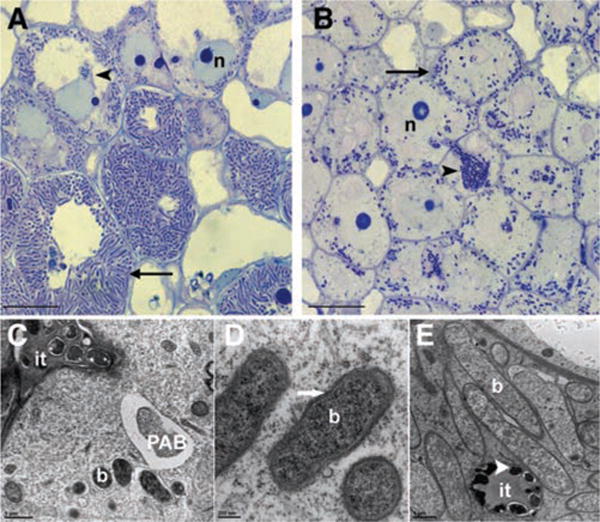
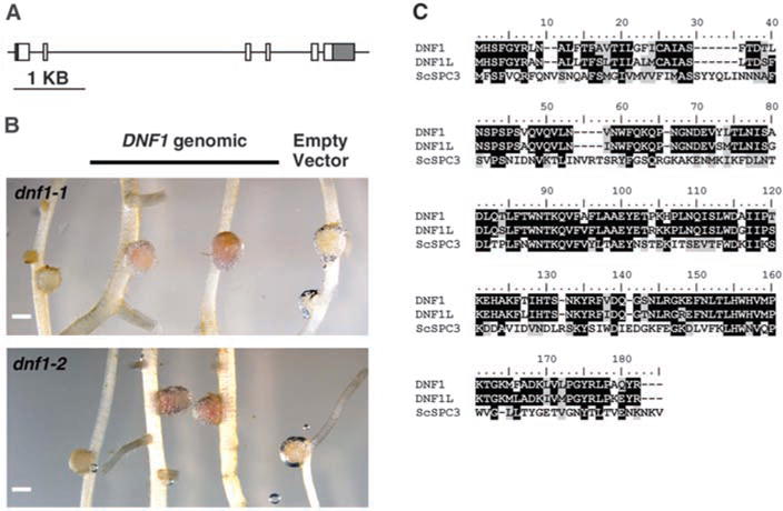
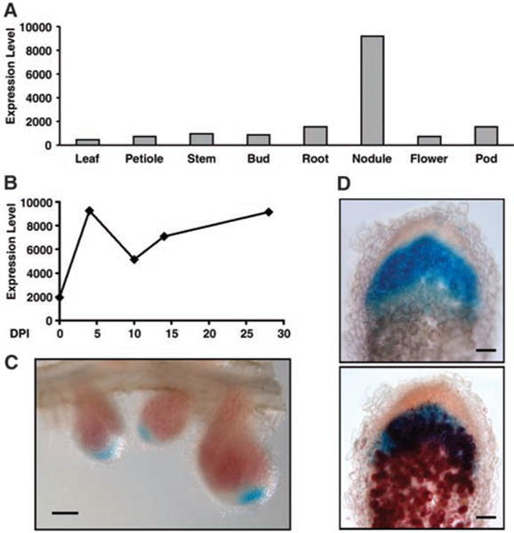
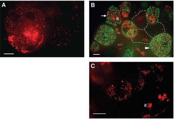
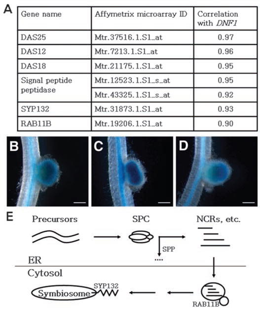
The nitrogen-fixing symbiosis between Sinorhizobium meliloti and its leguminous host plant Medicago truncatula occurs in a specialized root organ called the nodule. Bacteria that are released into plant cells are surrounded by a unique plant membrane compartment termed a symbiosome. We found that in the symbiosis-defective dnf1 mutant of M. truncatula, bacteroid and symbiosome development are blocked. We identified the DNF1 gene as encoding a subunit of a signal peptidase complex that is highly expressed in nodules. By analyzing data from whole-genome expression analysis, we propose that correct symbiosome development in M. truncatula requires the orderly secretion of protein constituents through coordinated up-regulation of a nodule-specific pathway exemplified by DNF1.


Figures

Fig. 1
Fig. 1
Cytological characterization of dnf1 root nodules. (A and B) High-magnification light microscopic image of longitudinal sections of a WT (A) and dnf1 (B) nodule 12 dpi, showing that, in all layers of the central tissue, the bacteroids in dnf1 are arrested at stage 1 (20). In WT, after release from the infection thread (it, arrowhead), the bacteria soon differentiate into elongated bacteroids (arrow). In dnf1, although many bacteroids are visible in the central part of the nodule, they have a size similar to that of the bacteria that are present in the infection threads. n, nucleus. (C) Transmission electron microscope (TEM) image of a freshly infected dnf1 nodule cell showing the infection thread (it), several bacteroids surrounded by a symbiosome membrane (b), and a pre-autophagic body (PAB) containing cytoplasm. (D) TEM image of an infected dnf1 nodule cell containing symbiosomes that remain arrested at stage 1. Arrows indicate symbiosome membrane. (E) Mature bacteroids in a WT nodule. These bacteroids are markedly bigger than the bacteria that are present in the it (arrowhead). Scale bars in (A) and (B), 10 μm; (C), 1 μm; (D), 200 nm; (E), 1 μm.
Fig. 2
Fig. 2
DNF1 encodes a subunit of the SPC. (A) Structure of the candidate DNF1 gene. Boxes represent exons, and the lines between them are introns. 5′ and 3′ untranslated regions are shaded. (B) Complementation of mutant nodule phenotype by introducing the genomic fragment of the predicted DNF1 gene. Pink color indicates the presence of leghemoglobin. Scale bars, 1 mm. (C) Alignment of protein sequences between DNF1, DNF1L of M. truncatula, and the Sa. cerevisiae homolog ScSPC3.
Fig. 3
Fig. 3
DNF1 expression pattern. (A) Expression level of DNF1 in various tissues. (B) Temporal profile of DNF1 expression in nodule. (C) Spatial pattern of DNF1 expression revealed by a promoter::GUS construct in 14-day-old nodules. Nodule samples were stained briefly to preserve the color of leghemoglobin. (D) DNF1 promoter activity in nodules inoculated with Rm1021 carrying hemA::lacZ. A DNF1 promoter::GUS transgenic nodule was stained for GUS activity, photographed (top), briefly fixed, sectioned manually, and then stained with Salmon-gal (bottom). Values in (A) and (B) are levels of Affymetrix probe signal based on microarray data from the M. truncatula Gene Expression Atlas. DPI: days post inoculation. Scale bars in (C), 1 mm; (D), 100 mm.
Fig. 4
Fig. 4
Localization of DNF1. Roots were inoculated with Rm1021 carrying hemA::mCherry, nodules were hand sectioned at 14 dpi, and the infection zones were imaged under a confocal microscope. (A) In WT nodule cells, bacteroids quickly become highly elongated. (B) DNF1 was fused to GFP, expressed from its own promoter, and introduced into dnf1-2 roots. In uninvaded cells, three of which are outlined with dotted lines, no robust GFP signals were observed. In cells being invaded by bacteria (arrow), where the end of the it is discernible, DNF1-GFP fluorescence first appears. In cells where bacteroids are fully mature (arrowhead), DNF1-containing structures are numerous and appear elongated and closely associated with symbiosomes. (C) In dnf1-2 mutant nodule cells, bacteroids remain small and undifferentiated after release from the it. Green, DNF1-GFP; red, Rm1021 hemA::mCherry. Scale bars, 10 μm.
Fig. 5
Fig. 5
Coordinated up-regulation of a nodule-specific protein secretory pathway. (A) Correlation in gene expression between DNF1 and additional components of the protein secretory pathway. Correlation values are provided by the M. truncatula Gene Expression Atlas (12) based on microarray data on vegetative and reproductive tissues, seed development, nodulation, mycorrhization, and chemical treatments. The signal peptide peptidase gene is represented by two probe sets. (B to D) Promoter::GUS activity in the nodule of DNF1 and its associated SPC genes DAS12 and DAS25. Transgenic roots were inoculated with Rm1021, and nodules were photographed at 14 dpi. (B) pDNF1::GUS; (C) pDAS12::GUS; and (D) pDAS25::GUS. Background GUS activity in the vasculature confirmed the presence of the transgene. In (C), a nodule with double primodia is being formed. (E) A model for a nodule-specific pathway dedicated to secrete protein constitutes toward the developing symbiosome. SPC denotes the DNF1-containing SPC, SPP refers to the signal peptide peptidase listed in (A), and NCRs are mature peptides from precursor proteins processed by SPC and SPP. Scale bar, 1 μm.

Similar articles

Cited by

References

    1. Starker CG, Parra-Colmenares AL, Smith L, Mitra RM, Long SR. Plant Physiol. 2006;140:671. - PMC - PubMed
    1. Mitra RM, Long SR. Plant Physiol. 2004;134:595. - PMC - PubMed
    1. Vasse J, de Billy F, Camut S, Truchet G. J Bacteriol. 1990;172:4295. - PMC - PubMed
    1. Thompson AR, Vierstra RD. Curr Opin Plant Biol. 2005;8:165. - PubMed
    1. Limpens E, et al. Plant Cell. 2009;21:2811. - PMC - PubMed

Publication types

MeSH terms