. 2010 Apr 16;106(7):1253-64.
doi: 10.1161/CIRCRESAHA.109.213116. Epub 2010 Feb 25.

Upregulation of Nox4 by hypertrophic stimuli promotes apoptosis and mitochondrial dysfunction in cardiac myocytes

Affiliations

Upregulation of Nox4 by hypertrophic stimuli promotes apoptosis and mitochondrial dysfunction in cardiac myocytes

Tetsuro Ago et al. Circ Res. .

Abstract

Rationale: NADPH oxidases are a major source of superoxide (O(2)(-)) in the cardiovascular system. The function of Nox4, a member of the Nox family of NADPH oxidases, in the heart is poorly understood.

Objective: The goal of this study was to elucidate the role of Nox4 in mediating oxidative stress and growth/death in the heart.

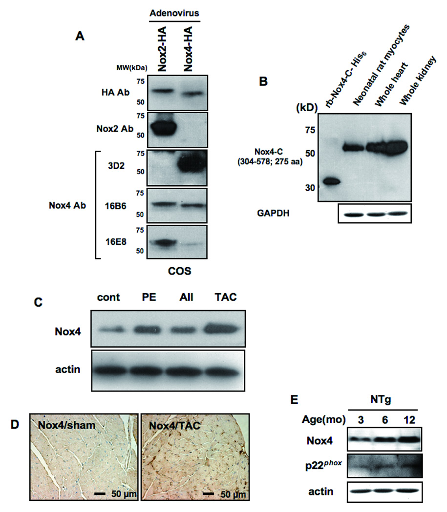
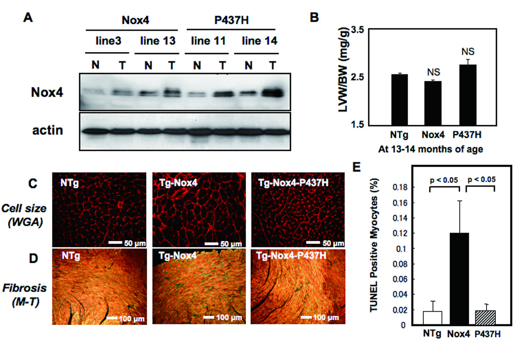
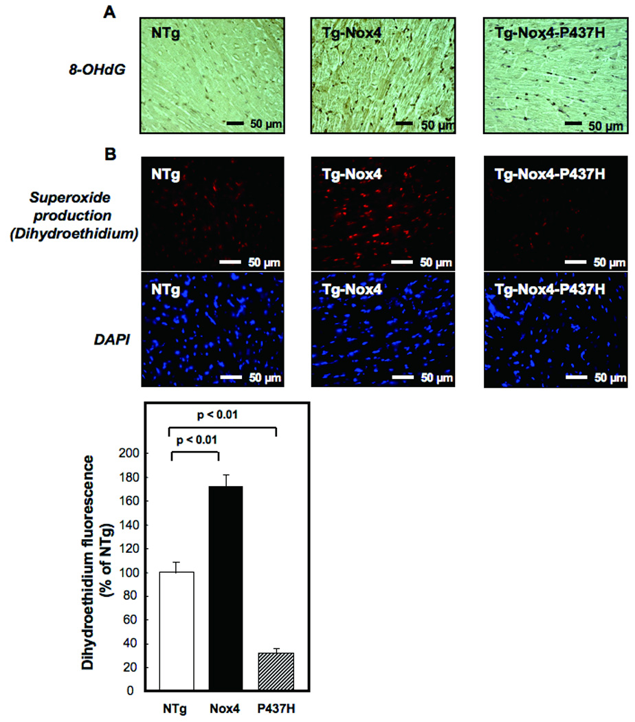
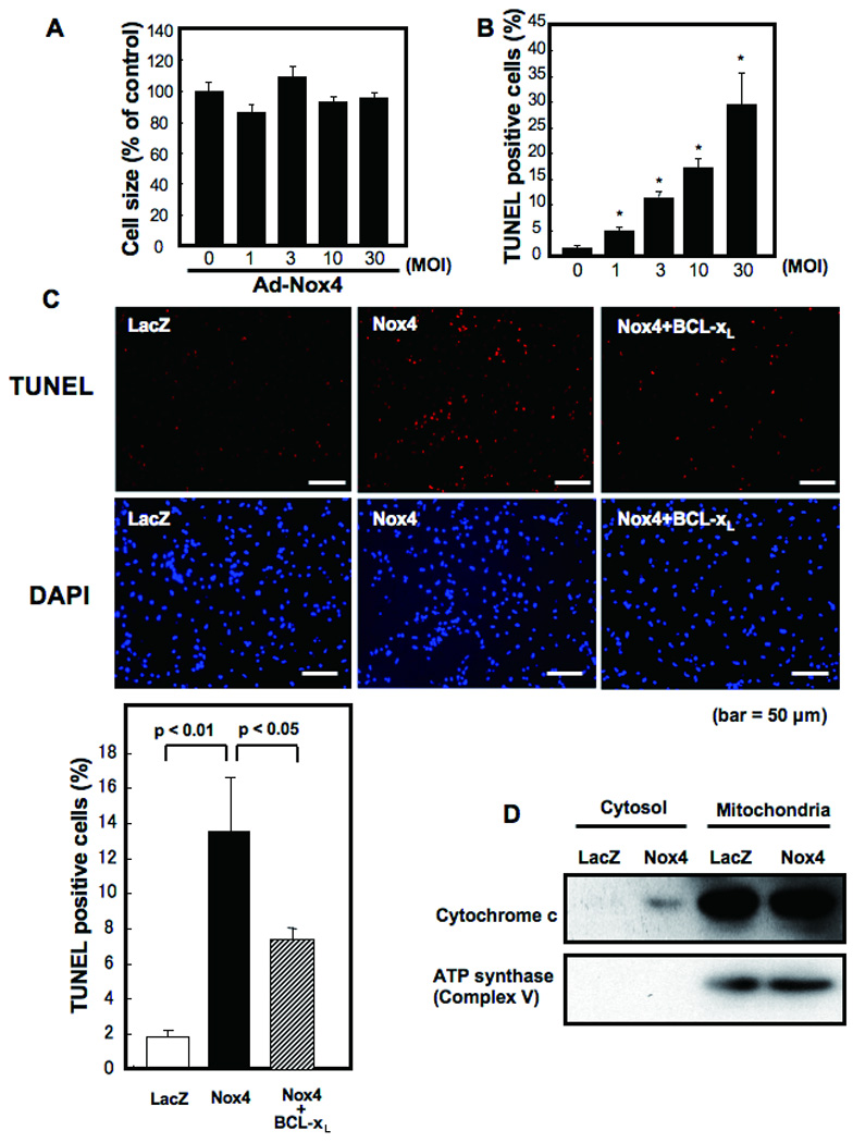
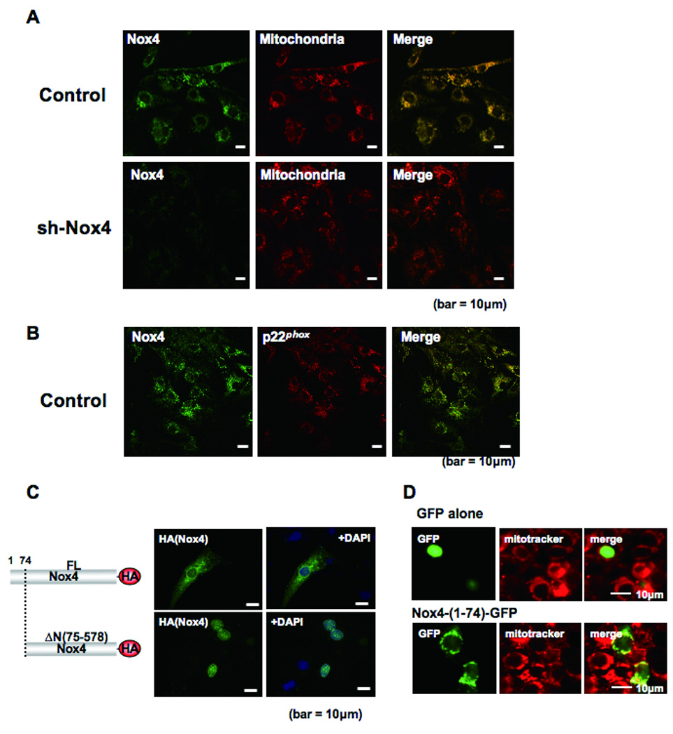
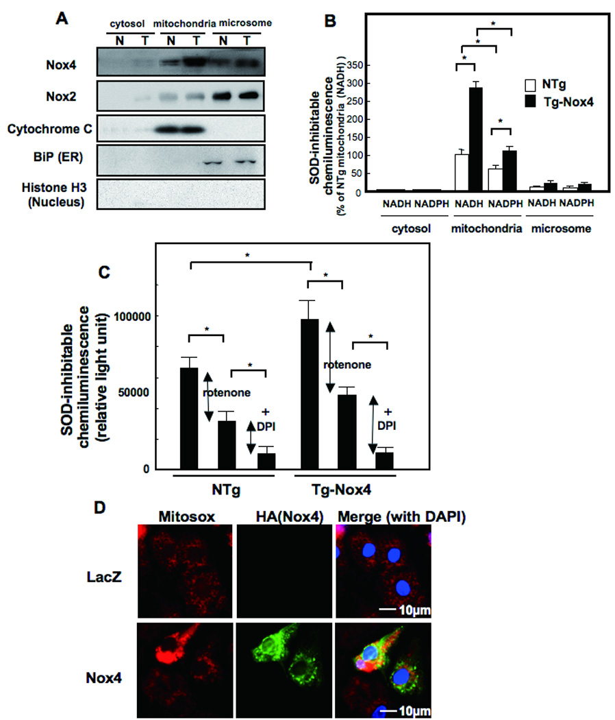
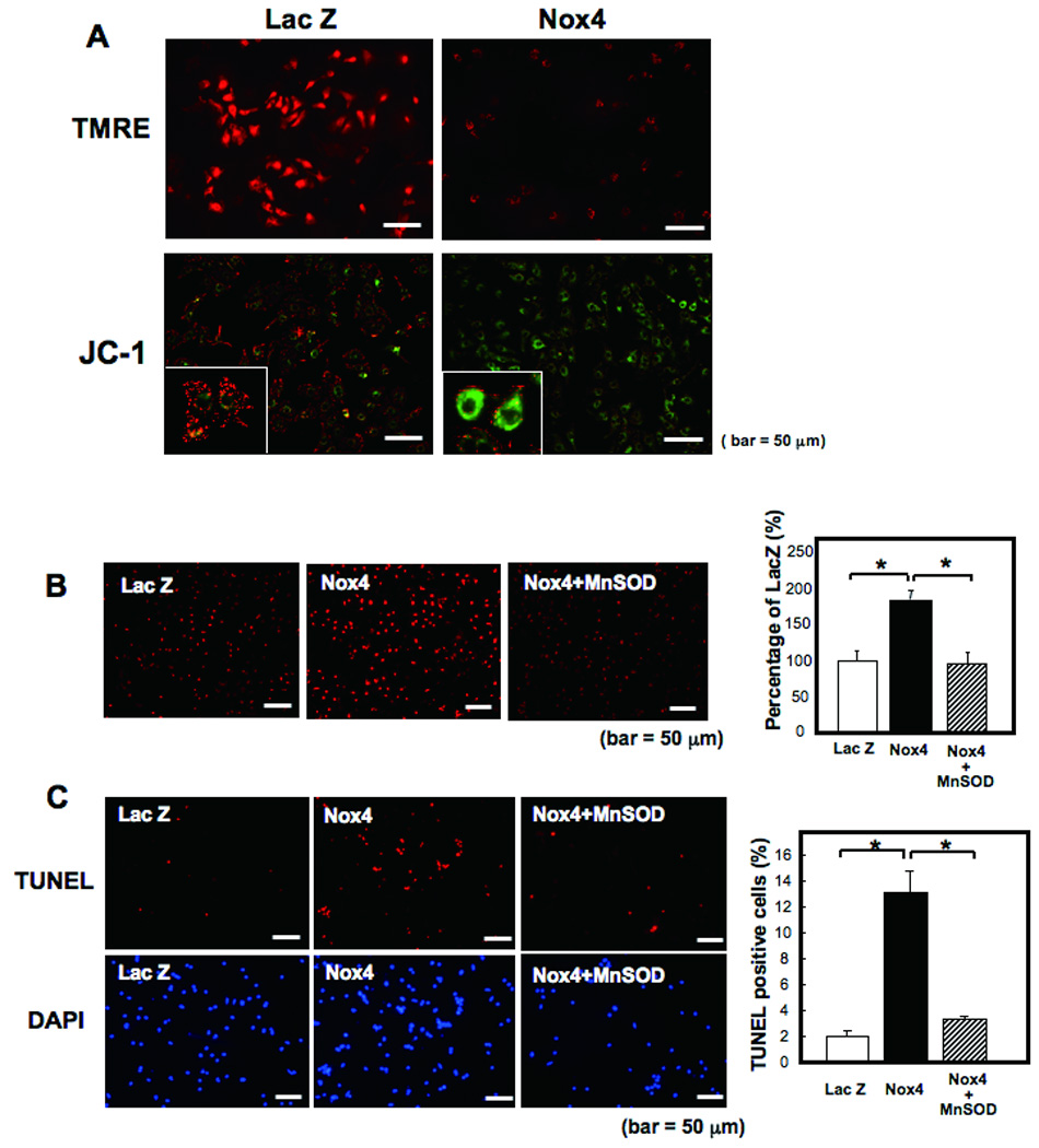
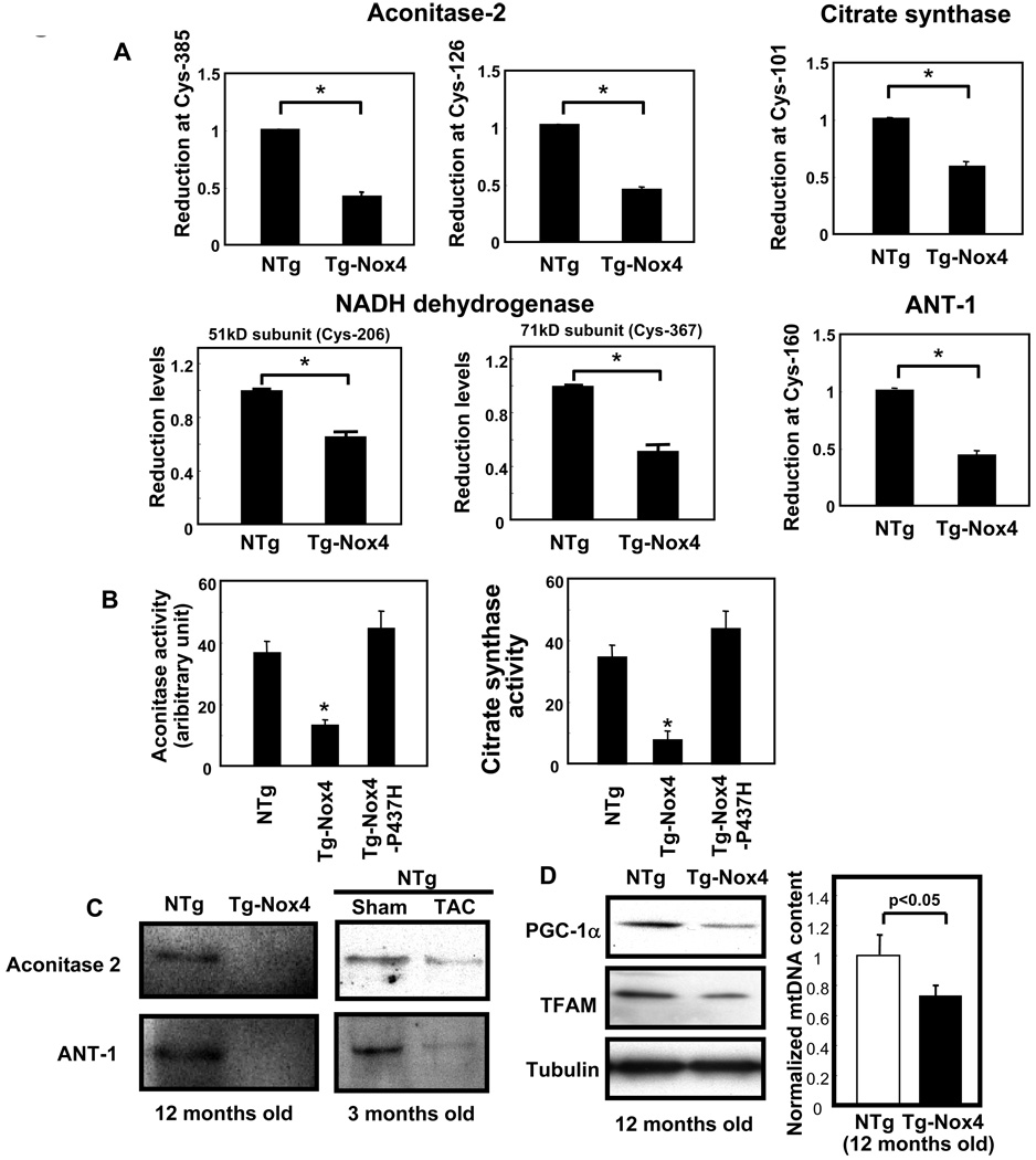
Methods and results: Expression of Nox4 in the heart was increased in response to hypertrophic stimuli and aging. Neither transgenic mice with cardiac specific overexpression of Nox4 (Tg-Nox4) nor those with catalytically inactive Nox4 (Tg-Nox4-P437H) showed an obvious baseline cardiac phenotype at young ages. Tg-Nox4 gradually displayed decreased left ventricular (LV) function with enhanced O(2)(-) production in the heart, which was accompanied by increased apoptosis and fibrosis at 13 to 14 months of age. On the other hand, the level of oxidative stress was attenuated in Tg-Nox4-P437H. Although the size of cardiac myocytes was significantly greater in Tg-Nox4 than in nontransgenic, the LV weight/tibial length was not significantly altered in Tg-Nox4 mice. Overexpression of Nox4 in cultured cardiac myocytes induced apoptotic cell death but not hypertrophy. Nox4 is primarily localized in mitochondria and upregulation of Nox4 enhanced both rotenone- and diphenyleneiodonium-sensitive O(2)(-) production in mitochondria. Cysteine residues in mitochondrial proteins, including aconitase and NADH dehydrogenases, were oxidized and their activities decreased in Tg-Nox4.

Conclusions: Upregulation of Nox4 by hypertrophic stimuli and aging induces oxidative stress, apoptosis and LV dysfunction, in part because of mitochondrial insufficiency caused by increased O(2)(-) production and consequent cysteine oxidation in mitochondrial proteins.


Figures

Figure 1
Figure 1
A) COS7 cells were transduced with adenovirus harboring either Nox2-HA or Nox4-HA. Immunoblot analyses were conducted with the indicated antibodies. Although Nox4 has a greater molecular weight than Nox2, it migrated faster on SDS-PAGE gel, possibly due to cleavage at the N-terminal mitochondrial localization signal (MLS). B) An immunoblot showing expression of Nox4 in the heart and cardiac myocytes. Recombinant protein used as an antigen for 3D2 and mouse kidney homogenate were used as positive controls. C) The effect of hypertrophic stimuli on expression of Nox4 in the mouse heart. Mice were subjected to continuous infusion with phenylephrine (PE) or angiotensin II (AII) or to transverse aortic constriction (TAC) for 2 weeks. N=4–5. D) Nox4 expression in the mouse heart subjected to sham or TAC. Immunostaining was conducted with anti-Nox4 antibody (3D2). E) The effect of aging upon Nox4, p22phox, and actin expression in the control mouse heart was evaluated by immunoblot analyses.
Figure 2
Figure 2
A) Expression of Nox4 in Tg-Nox4 and Tg-Nox4-P437H. N, non-transgenic; T, transgenic. B) Left ventricular weight/body weight (LVW/BW) at 13–14 months of age. NS, not significant vs NTg. N=8–9. (C–E) Age-dependent changes in histology in Tg-Nox4, Tg-Nox4-P437H and NTg. C) LV myocyte cross sectional area as evaluated by WGA staining, D) LV fibrosis as determined by Masson Trichrome staining, and E) apoptosis as determined by TUNEL staining.
Figure 3
Figure 3
The extent of oxidative stress in the LV was determined by 8-hydroxyl-deoxyguanosine (8-OHdG) staining (A) and dihydroethidium (DHE) staining (B). In B, DAPI staining shows nuclei. LV sections were prepared from Tg-Nox4, Tg-Nox4-P437H and NTg mice.
Figure 4
Figure 4
Cultured neonatal rat cardiac myocytes were transduced with Ad-Nox4 or Ad-LacZ at various multiplicities of infection (MOI) for 48 hours. Data are from 4–6 experiments. (A) Mean cell size of control cardiac myocytes is expressed as 100%. Changes were not significant. (B) The number of apoptotic cells was evaluated with TUNEL staining. *p<0.05 vs control (0 MOI). (C) Cultured cardiac myocytes were transduced with Ad-LacZ, Ad-Nox4, or Ad-Nox4 + Ad-Bcl-xL. Representative TUNEL and DAPI staining and the results of the quantitative analysis are shown. (D) Cultured cardiac myocytes were transduced with Ad-LacZ or Ad-Nox4. Cytosolic and mitochondrial fractions were isolated and expression of cytochrome c and F0F1 ATP synthase was evaluated with immunoblot analyses.
Figure 5
Figure 5
Subcellular localization of Nox4 in cardiac myocytes. A) Cultured cardiac myocytes were transduced with adenovirus harboring shRNA-scramble or shRNA-Nox4. Myocytes were co-stained with anti-Nox4 antibody and anti-F0F1 ATP synthase antibody, a marker of mitochondria. Merged images are shown on the right. B) Cultured cardiac myocytes were co-stained with anti-Nox4 and anti-p22phox antibodies. A merged image is shown on the right. C) Cultured cardiac myocytes were transduced with an expression plasmid harboring full length (FL) Nox4-HA or Nox4 lacking MLS (ΔN(75–578))-HA. Forty-eight hours after transfection, cells were stained with anti-HA antibody and DAPI. Truncation of the N-terminal region (1–74) causes disappearance of the perinuclear staining of Nox4 in cardiac myocytes. D) Cardiac myocytes were transduced with expression plasmids harboring either GFP alone or Nox4 (1–74)-GFP. Representative images of GFP, Mitotracker and merged images are shown.
Figure 6
Figure 6
Localization of Nox4 in cardiac myocytes. (A and B) Cytosolic, mitochondrial membrane, and microsomal fractions were prepared from aging NTg and Tg-Nox4 mouse hearts. (A) Immunoblot analyses were conducted with anti-Nox4 monoclonal antibody (3D2) and anti-Nox2 polyclonal antibody. The purity of the mitochondrial fraction was confirmed by the lack of histone H3 or BiP staining. The purity of cytosolic fraction was confirmed by the lack of cytochrome c. (B, C) NADH- (B, C) and NADPH-dependent (B) O2 release was determined by the lucigenin method. The SOD inhibitable component of O2 release from each fraction was determined. *p<0.05. (C) NADH-dependent and SOD inhibitable O2 release from the mitochondrial membrane fraction consists of Rotenone-sensitive (shown by red arrow) and Rotenone-insensitive, DPI-sensitive (shown by blue arrow) components. The Rotenone-insensitive, DPI-sensitive components may include O2 release from Nox localized in the mitochondria *p<0.05. (D) Cultured neonatal rat cardiac myocytes were transduced with adenovirus harboring LacZ or Nox4. The extent of oxidative stress in the mitochondria was evaluated using MitoSOX™.
Figure 7
Figure 7
A) Cultured neonatal rat cardiac myocytes were transduced with adenovirus harboring Nox4 or Lac Z. After 48 hours, the cells were subjected to TMRE and JC-1 staining for mitochondrial membrane potential assessment. Note that red staining indicates polarized mitochondria in TMRE and JC-1 staining. Green staining indicates depolarized mitochondria in JC-1 staining. B,C) Cultured neonatal rat cardiac myocytes were transduced with adenovirus harboring Nox4 or Lac Z, or co-transduced with Ad-Nox4 and Ad-MnSOD. B) After 48 hours, superoxide production was evaluated with DHE staining. *p<0.05. C) The number of apoptotic cells were evaluated with TUNEL staining. *p<0.05.
Figure 8
Figure 8
Overexpression of Nox4 causes mitochondrial dysfunction. Purified mitochondrial fractions were prepared from aging Tg-Nox4 and NTg mouse hearts and subjected to ICAT proteomics or biochemical assays. A) The ICAT signal at cysteine 385 of aconitase, cysteine 126 of aconitase, cysteine 101 of citrate synthase, cysteine 206 of NADH dehydrogenase (51 kD subunit), cysteine 367 of NADH dehydrogenase (71 kD subunit), and cysteine 160 of adenine nucleotide translocase type 1 (ANT1) were significantly lower in Tg-Nox4 hearts than in NTg hearts, suggesting that these cysteines are oxidized. (B) The aconitase activity and the citrate synthase activity in the heart were compared among Tg-Nox4, Tg-Nox4-P437H and NTg mice. C) Twelve month old NTg and Tg-Nox4 mouse hearts were lysed in the presence of biotinylated iodoacetamide. Biotinylated proteins were pulled down on streptavidin beads and subjected to immunoblotting (Left panel). Three month old NTg mice were subjected to TAC or sham operation. The hearts were treated in the same way as above (Right panel). D) Evaluation of mitochondrial biogenesis. Twelve month old NTg or Tg-Nox4 mouse hearts were subjected to immunoblotting using antibodies raised against PGC-1α and TFAM (Left panel). Quantitative real-time PCR for mitochondrial DNA (Right panel). *p<0.05 vs NTg.

References

    1. Liu H, Colavitti R, Rovira II, Finkel T. Redox-dependent transcriptional regulation. Circ Res. 2005;97:967–974. - PubMed
    1. Sorescu D, Griendling KK. Reactive oxygen species, mitochondria, and NAD(P)H oxidases in the development and progression of heart failure. Congest Heart Fail. 2002;8:132–140. - PubMed
    1. Tsutsui H, Kinugawa S, Matsushima S. Mitochondrial oxidative stress and dysfunction in myocardial remodelling. Cardiovasc Res. 2009;81:449–456. - PubMed
    1. Ide T, Tsutsui H, Hayashidani S, Kang D, Suematsu N, Nakamura K, Utsumi H, Hamasaki N, Takeshita A. Mitochondrial DNA damage and dysfunction associated with oxidative stress in failing hearts after myocardial infarction. Circ Res. 2001;88:529–535. - PubMed
    1. Balaban RS, Nemoto S, Finkel T. Mitochondria, oxidants, and aging. Cell. 2005;120:483–495. - PubMed

Publication types

MeSH terms