. 2010 May;59(5):1202-10.
doi: 10.2337/db09-1177. Epub 2010 Feb 25.

Unique arrangement of alpha- and beta-cells in human islets of Langerhans

Affiliations

Unique arrangement of alpha- and beta-cells in human islets of Langerhans

Domenico Bosco et al. Diabetes. 2010 May.

Abstract

Objective: It is generally admitted that the endocrine cell organization in human islets is different from that of rodent islets. However, a clear description of human islet architecture has not yet been reported. The aim of this work was to describe our observations on the arrangement of human islet cells.

Research design and methods: Human pancreas specimens and isolated islets were processed for histology. Sections were analyzed by fluorescence microscopy after immunostaining for islet hormones and endothelial cells.

Results: In small human islets (40-60 mum in diameter), beta-cells had a core position, alpha-cells had a mantle position, and vessels laid at their periphery. In bigger islets, alpha-cells had a similar mantle position but were found also along vessels that penetrate and branch inside the islets. As a consequence of this organization, the ratio of beta-cells to alpha-cells was constantly higher in the core than in the mantle part of the islets, and decreased with increasing islet diameter. This core-mantle segregation of islet cells was also observed in type 2 diabetic donors but not in cultured isolated islets. Three-dimensional analysis revealed that islet cells were in fact organized into trilaminar epithelial plates, folded with different degrees of complexity and bordered by vessels on both sides. In epithelial plates, most beta-cells were located in a central position but frequently showed cytoplasmic extensions between outlying non-beta-cells.

Conclusions: Human islets have a unique architecture allowing all endocrine cells to be adjacent to blood vessels and favoring heterologous contacts between beta- and alpha-cells, while permitting homologous contacts between beta-cells.


Figures

FIG. 1.
FIG. 1.
Organization of α- and β-cells in human pancreatic islets. Sections of human pancreata with islets of different sizes were either double-labeled for insulin (red) and glucagon (green) (A–C) or triple-labeled for insulin (blue), glucagon (red), and CD34 (green) (G–I). D–F and J–L: Outlines of endocrine tissue labeled for insulin and glucagon were drawn. Except for the 40- to 60-μm–diameter islets, all islets displayed one or several unstained empty areas (vascular channels) at their core. Most glucagon-expressing cells were located around vascular channels and at the mantle of islets, independent of their size. Insulin-expressing cells seemed clustered into discrete ovoid areas surrounded by α-cells. In triple-labeled sections (G–I), vascular channels displayed staining for CD34, indicating that they contained vessels. Islets shown are representative of at least 200 islets observed on sections from 16 different pancreata. Scale bars, 50 μm. (A high-quality digital representation of this figure is available in the online issue.)
FIG. 2.
FIG. 2.
Distribution of α- and β-cells according to different defined islet subregions. A: Pancreatic islet labeled by immunofluorescence for insulin (red) and glucagon (green); rims (white lines) delimiting mantle and core subregions and one vascular channel area are shown; scale bar, 10 μm. After immunofluorescence, areas labeled for insulin (b) and glucagon (a) were measured and results expressed as b/(a+b) ratio. This ratio was calculated for areas measured in the different subregions (mantle, core, and vessels) and in the whole islets (whole). Analyses were performed on histologic sections from nondiabetic human (B), type 2 diabetic human (C), and control mouse (E) pancreata and cultured isolated human islets (D). Columns are means ± SEM. B: n = 193 islets from 16 pancreata. C: n = 54 islets from five pancreata. D: n = 20 islets from two pancreata. E: n = 50 islets from one pancreas. ND, not determined. (A high-quality digital representation of this figure is available in the online issue.)
FIG. 3.
FIG. 3.
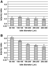
Distribution of α- and β-cells according to islet apparent size. Islets from human pancreas sections analyzed in Fig. 2 are shown here according to their apparent diameter. After immunofluorescence, insulin- and glucagon-labeled areas were measured and results expressed as b/(a+b) ratio, where a and b were representing glucagon and insulin labeled areas, respectively. A: b/(a+b) ratio calculated for areas measured in the 20-μm mantle part of islets. B: b/(a+b) ratio calculated for areas measured in the core of the islets. Columns are means ± SEM. n = 39 for islets with 50- to 100-μm diameter, 60 for islets with 100- to 150-μm diameter, 58 for islets with 150- to 200-μm diameter, 24 for islets with 200- to 250-μm diameter, and 12 for islets with 250- to 300-μm diameter.
FIG. 4.
FIG. 4.
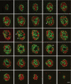
Three-dimensional analysis of human pancreatic islets. Consecutive sections through an entire pancreatic islet labeled for insulin (red) and glucagon (green). Images show that insulin (red)– and glucagon (green)–stained cells are organized into continuous 3-D networks that span the entire islet. Sections at both ends show islet profiles with an apparent similar core-mantle structure to that of 40- to 60-μm–diameter islets. These consecutive images also reveal that vascular channels were in fact continuous ramified structures that were connected in places with the surrounding islet tissue (arrowheads). Image series is representative of 15 different pancreatic islets analyzed from one pancreas. Scale bar, 50 μm. (A high-quality digital representation of this figure is available in the online issue.)
FIG. 5.
FIG. 5.
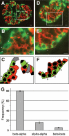
Unique association between α- and β-cells in pancreatic islets. Pancreatic islets labeled for glucagon (green) and insulin (red) are shown at low magnification (A and D). Boxed areas are shown at higher magnification in B and E. α- and β-cells lining vascular channels in B are depicted in C. α- and β-cells lining the islet mantle in E are depicted in F. Arrowheads point to cytoplasmic extensions of β-cells that span α-cells. Scale bars, 10 μm in A and D, and 5 μm in B and E. Heterologous contacts between β- and α-cells (beta-alpha) and homologous contacts between α-cells (alpha-alpha) and β-cells (beta-beta) around empty areas were scored. Their relative frequencies are shown in G. Columns are means ± SEM; n = 52 islets from 10 pancreata. (A high-quality digital representation of this figure is available in the online issue.)
FIG. 6.
FIG. 6.
Unique association between α- and β-cells in cultured islet cells. Human islet cells were isolated and cultured for 24 h. After a double immunofluorescence for insulin (red) and glucagon (green), islet cells were analyzed by confocal microscopy. A–C: Images showing a cell pair composed of one α-cell (A) surrounded by one β-cell (B); the merged image is shown in C. The cell pair shown here is representative of cell pairs observed in different human cell preparations from at least 10 different pancreata. D: One series of consecutive merged images of a cell pair composed of one α-cell (green) surrounded by a β-cell (red). Scale bars, 10 μm. E: All heterologous contacts between α- and β-cells were scored according to their type of association: a β-cell wrapping an α-cell (beta wrapping alpha), neutral apposition between α- and β-cells (alpha-beta), and an α-cell wrapping a β-cell (alpha wrapping beta). Results are shown as relative frequencies and columns are means ± SEM of five islet cell preparations from five different pancreata. From all heterogeneous contacts between α- and β-cells, the percentage of α-cells whose profile was round and perimeter almost completely wrapped by a β-cell as in D was 38 ± 8 (means ± SEM of three experiments). (A high-quality digital representation of this figure is available in the online issue.)
FIG. 7.
FIG. 7.
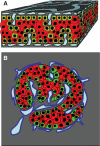
Model of endocrine cell and vessel organization in human islets. A: α-Cells (green) and β-cells (red) are organized into a thick folded plate lined at both sides with vessels (blue). α-Cells are mostly at the periphery of the plate and in close contact with vessels. β-Cells occupy a more central part of the plate and most of them develop cytoplasmic extension that runs between α-cells and reaches the surface of vessels. B: The plate with adjacent vessels is folded so that it forms an islet.

Similar articles

Cited by

References

    1. Orci L, Unger RH. Functional subdivision of islets of Langerhans and possible role of D cells. Lancet 1975;2:1243–1244 - PubMed
    1. Gannon M, Ray MK, Van Zee K, Rausa F, Costa RH, Wright CV. Persistent expression of HNF6 in islet endocrine cells causes disrupted islet architecture and loss of beta cell function. Development 2000;127:2883–2895 - PubMed
    1. Bosco D, Orci L, Meda P. Homologous but not heterologous contact increases the insulin secretion of individual pancreatic B-cells. Exp Cell Res 1989;184:72–80 - PubMed
    1. Samols E, Stagner JI. Intra-islet regulation. Am J Med 1988;85:31–35 - PubMed
    1. Samols E, Stagner JI. Islet somatostatin: microvascular, paracrine, and pulsatile regulation. Metabolism 1990;39:55–60 - PubMed

Publication types