. 2010 Apr 23;285(17):12971-8.
doi: 10.1074/jbc.M109.076091. Epub 2010 Feb 25.

TRAF3 controls activation of the canonical and alternative NFkappaB by the lymphotoxin beta receptor

Affiliations

TRAF3 controls activation of the canonical and alternative NFkappaB by the lymphotoxin beta receptor

Pradeep Bista et al. J Biol Chem. .

Abstract

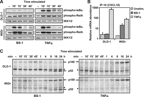
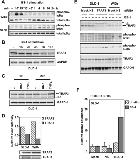
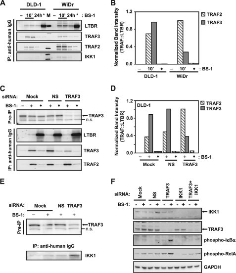
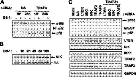
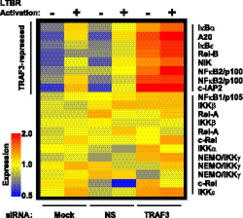
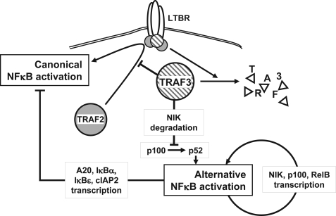
Components of lymphotoxin beta receptor (LTBR)-associated signaling complexes, including TRAF2, TRAF3, NIK, IKK1, and IKK2 have been shown to participate in the coupling of LTBR to NFkappaB. Here, we report that TRAF3 functions as a negative regulator of LTBR signaling via both canonical and non-canonical NFkappaB pathways by two distinct mechanisms. Analysis of NFkappaB signaling in cell lines with functionally intact NFkappaB pathway but lacking LTBR-mediated induction of NFkappaB target genes revealed an inverse association of cellular TRAF3 levels with LTBR-specific defect in canonical NFkappaB activation. Increased expression of TRAF3 correlated with its increased recruitment to LTBR-induced signaling complexes, decreased recruitment of TRAF2, and attenuated phosphorylation of IkappaB alpha and RelA. In contrast, activation of NFkappaB by TNF did not depend on TRAF3 levels. siRNA-mediated depletion of TRAF3 promoted recruitment of TRAF2 and IKK1 to activated LTBR, enabling LTBR-inducible canonical NFkappaB signaling and NFkappaB target gene expression. TRAF3 knock-down also increased mRNA and protein expression of several non-canonical NFkappaB components, including NFkappaB2/p100, RelB, and NIK, accompanied by processing of NFkappaB2/p100 into p52. These effects of TRAF3 depletion did not require LTBR signaling and were consistent with autonomous activation of the non-canonical NFkappaB pathway. Our data illustrate the function of TRAF3 as a dual-mode repressor of LTBR signaling that controls activation of canonical NFkappaB, and de-repression of the intrinsic activity of non-canonical NFkappaB. Modulation of cellular TRAF3 levels may thus contribute to regulation of NFkappaB-dependent gene expression by LTBR by affecting the balance of LTBR-dependent activation of canonical and non-canonical NFkappaB pathways.


Figures

FIGURE 1.
FIGURE 1.
LTBR-specific activation of canonical NFκB is uncoupled in certain cells and correlates with differential cytokine gene activation. DLD-1 and WiDr cells were treated with agonist LTBR antibody (BS-1) at 100 ng/ml, or TNFα (20 ng/ml) for indicated times. Cell lysates were analyzed by Western blot for (A) phosphorylated IκBα (Ser-32/Ser-36), phosphorylated RelA (Ser-536), or IKK1/2 (to control for loading), and (C) NFκB2 (*, nonspecific bands). B, RNA isolated from untreated cells (▧), cells treated with BS-1 (■), or with TNFα (▤) for 4 h were analyzed by real-time qPCR for IP-10 transcripts. The data are shown as housekeeping gene (GAPDH)-normalized values from quadruplicate samples (average + S.D.).
FIGURE 2.
FIGURE 2.
TRAF3 inhibits LTBR-induced canonical NFκB activation. A, cells were treated with 100 ng/ml agonist LΤΒR antibody (BS-1) for indicated times, and the lysates were analyzed by Western blot for phosphorylated IκBα (Ser-32/Ser-36) and total IκBα. B, samples from BS-1 stimulated DLD-1 cells were also probed for TRAF3 or GAPDH (loading control). C, DLD-1 cells were left untreated, or treated for 10 min and 20 h with 100 ng/ml BS-1, or 20 ng/ml TNFα, and lysates analyzed for TRAF3 and GAPDH (loading control) by Western blot (n.s., nonspecific bands). D, total lysates from untreated DLD-1 and WiDr cells were analyzed by Western blots for TRAF2 and TRAF3, and band intensities were quantified by densitometry. E, cells were mock transfected (Mock), or transfected with either a nonspecific control siRNA (NS), or TRAF3 siRNA for 48 h. The cells were then left untreated (− lanes), or treated (+ lanes) with BS-1 for 10 min, and samples analyzed by Western blots. Blots were probed for different proteins as indicated (n.s., nonspecific band). F, DLD-1 cells were transfected as in C, and left untreated (unstim.) or stimulated with BS-1 for 4 h. RNA was collected and analyzed by real-time qPCR for IP-10 transcripts (unstimulated: ▧; BS-1 treated: ■). Results are normalized to GAPDH transcripts and shown as average + S.D. from quadruplicate samples.
FIGURE 3.
FIGURE 3.
Cellular TRAF3 level controls the composition of LTBR signaling complexes. A, cells were left untreated (− lanes), or treated for 10 min (10′ lanes) and 24 h (24 h lanes) with 100 ng/ml agonist LTBR antibody (BS-1). For positive controls, cells were lysed first and BS-1 added to the unstimulated lysates (*, lanes). BS-1-bound complexes were immunoprecipitated using anti-human IgG conjugated agarose beads, and analyzed by Western blots for LTBR, TRAF3, TRAF2, and IKK1, as indicated. B, band intensities were analyzed by densitometry, and data shown as ratio of either TRAF2 (▧), or TRAF3 (■), to LTBR. C, DLD-1 cells were mock transfected or transfected with non-silencing control siRNA (NS), or siRNA directed against TRAF3, and treated as in A. Pre-IP samples were probed for TRAF3 (n.s., nonspecific band), and immunoprecipitates were analyzed as in A for LTBR, TRAF2, and TRAF3. D, bands from C were quantified by densitometry as described in B. E, DLD-1 cells were treated as in C, lysates were probed for TRAF3 levels before immunoprecipitation to ensure knock-down, and immunoprecipitates were analyzed for IKK1 by Western blot. F, DLD-1 cells were transfected with siRNA against TRAF3 and IKK1, alone or in combination. 48 h after transfection, cells were left unstimulated or stimulated with BS-1 for 10 min and lysates analyzed by Western blots for IKK1, TRAF3, phospho-IκBα (Ser-32/Ser-36), phospho-RelA (Ser-536), and GAPDH (to control for loading).
FIGURE 4.
FIGURE 4.
TRAF3 knock-down leads to constitutive, NIK-dependent processing of NFκB2/p100. A, DLD-1 cells transfected with a non-silencing control siRNA (NS) or TRAF3 siRNA for 48 h were left untreated (− lanes) or treated (+ lanes) with 100 ng/ml anti-LTBR antibody (BS-1) for 10 min and 24 h. Lysates were analyzed by Western blot for NFκB2 (p100, and processed p52 are indicated by arrows). B, DLD-1 cells treated for indicated times with BS-1 were analyzed by Western blot for NIK (indicated by an arrow; n.s., nonspecific band). C, DLD-1 cells were mock transfected (Mock), transfected with non-silencing control siRNA (NS), or with siRNA against indicated targets either alone or in combination with TRAF3 siRNA. Cells were cultured for 48 h after transfection in the absence of any stimulus, and samples were probed for the indicated proteins by Western blots.
FIGURE 5.
FIGURE 5.
TRAF3 represses a group of NFκB-related genes. DLD-1 cells were mock transfected (Mock), or transfected with a non-silencing siRNA (NS) or TRAF3 siRNA. 48 h post-transfection, cells were left untreated (−), or treated (+) with agonist anti-LTBR antibody (BS-1) for 4 h. RNA was collected and profiled for NFκB-related transcripts by microarray.
FIGURE 6.
FIGURE 6.
Proposed model for NFκB repression by TRAF3 in LTBR activation. TRAF3 inhibits activation of the classical NFκB pathway by interfering with the formation of productive signaling complexes at LTBR. A high level of TRAF3 interferes with the recruitment of TRAF2 to the receptor. In parallel, TRAF3 induces NIK degradation and consequently inhibits the activation of the alternative arm of NFκB. During LTBR signaling, the alternative arm is activated as a result of TRAF3 degradation and NIK derepression. The sustained activation of the alternative arm in the absence of TRAF3 is maintained by an autoactivation loop. Alternative arm activation up-regulates NIK, NFκB2/p100, and RelB transcription by p52 and RelB, and the continued processing of NFκB2/p100 into p52 is induced by derepressed NIK. Additionally, persistent alternative arm activation up-regulates NFκB1 inhibitors such as IκBα and A20, keeping the classical NFκB pathway inactive in the absence of activation signals.

References

    1. Schneider K., Potter K. G., Ware C. F. (2004) Immunol. Rev. 202, 49–66 - PubMed
    1. Bonizzi G., Karin M. (2004) Trends Immunol. 25, 280–288 - PubMed
    1. Hayden M. S., Ghosh S. (2004) Genes Dev. 18, 2195–2224 - PubMed
    1. Beinke S., Ley S. C. (2004) Biochem. J. 382, 393–409 - PMC - PubMed
    1. Locksley R. M., Killeen N., Lenardo M. J. (2001) Cell 104, 487–501 - PubMed

MeSH terms