. 2010 Apr 23;285(17):13142-53.
doi: 10.1074/jbc.M110.114561. Epub 2010 Feb 25.

A role for huntington disease protein in dendritic RNA granules

Affiliations

A role for huntington disease protein in dendritic RNA granules

Jeffrey N Savas et al. J Biol Chem. .

Abstract

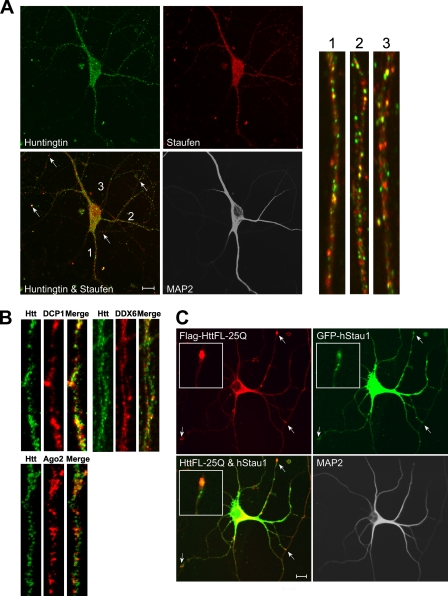
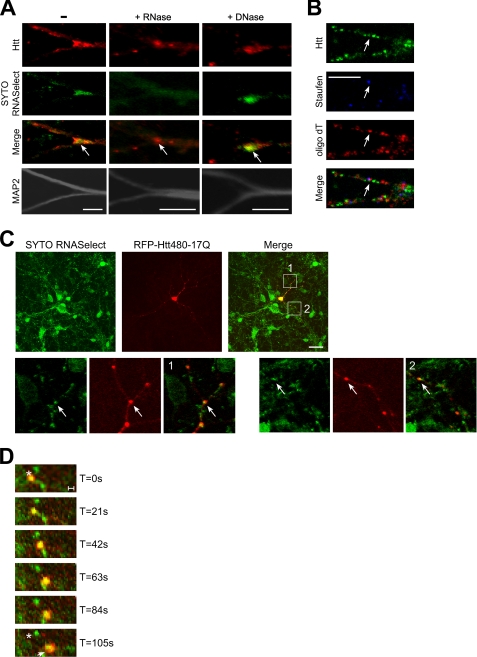
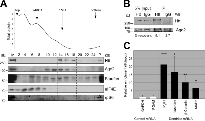
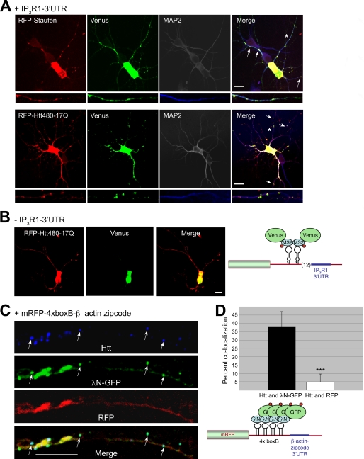
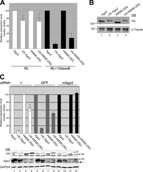
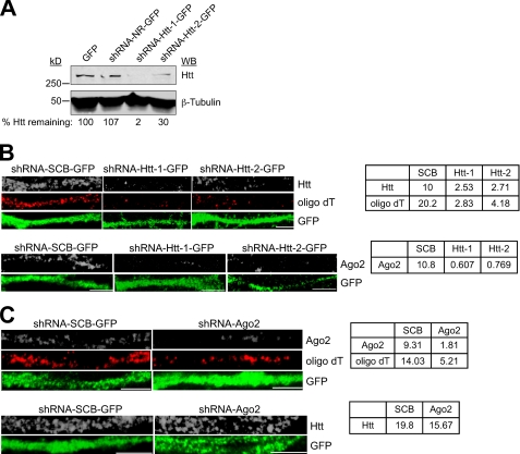
Regulated transport and local translation of mRNA in neurons are critical for modulating synaptic strength, maintaining proper neural circuitry, and establishing long term memory. Neuronal RNA granules are ribonucleoprotein particles that serve to transport mRNA along microtubules and control local protein synthesis in response to synaptic activity. Studies suggest that neuronal RNA granules share similar structures and functions with somatic P-bodies. We recently reported that the Huntington disease protein huntingtin (Htt) associates with Argonaute (Ago) and localizes to cytoplasmic P-bodies, which serve as sites of mRNA storage, degradation, and small RNA-mediated gene silencing. Here we report that wild-type Htt associates with Ago2 and components of neuronal granules and co-traffics with mRNA in dendrites. Htt was found to co-localize with RNA containing the 3'-untranslated region sequence of known dendritically targeted mRNAs. Knockdown of Htt in neurons caused altered localization of mRNA. When tethered to a reporter construct, Htt down-regulated reporter gene expression in a manner dependent on Ago2, suggesting that Htt may function to repress translation of mRNAs during transport in neuronal granules.


Figures

FIGURE 1.
FIGURE 1.
Huntingtin is present at a subset of neuronal granules in dendrites. A, rat hippocampal neurons (DIV 14) stained with α-Htt, α-Staufen (neuronal granule marker) and α-MAP2 (somatodendritic marker) antibodies (scale bar, 10 μm). Enlarged views of three primary dendrites stained with Htt and Staufen antibodies (labeled 1, 2, and 3; ∼40 μm is shown). B, hippocampal neurons stained with α-Htt, α-DCP1, α-DDX6, and α-Ago2 antibodies. ∼40 μm (upper panel) and 21 μm (lower panel) of primary dendrites are shown. C, cortical neuron (DIV 7) transfected with FLAG-HttFL-25Q and GFP-hStaufen1 and subsequently probed with α-FLAG and α-MAP2 antibodies. Scale bar, 10 μm. Inset, enlarged view of the area indicated by the arrow near the top of each image.
FIGURE 2.
FIGURE 2.
Huntingtin co-localizes with dendritic mRNA in rodent neurons. A, mouse hippocampal neurons (DIV 14) stained live with SYTO RNASelect, subsequently incubated with DNase or RNase where indicated, and lastly probed with α-Htt and α-MAP2 antibodies. Scale bar, 5 μm. B, mouse hippocampal neurons were probed with α-Htt or α-Staufen antibodies and oligo(dT) FISH. Scale bar, 5 μm. C, Rat E19 cortical neurons (DIV 5) transfected with Htt480-17Q were stained with SYTO RNASelect 24 h after transfection. Htt and RNA are shown in red and green, respectively. Enlarged areas 1 and 2 are shown in the lower panel. Scale bar (top, merge), 30 μm. D, merged time-lapse images of RFP-Htt480-17Q and SYTO RNASelect. *, initial location of RFP-Htt480-17Q, and the arrow shows the location after 105 s (see also supplemental Movie 1). Scale bar, 1 μm.
FIGURE 3.
FIGURE 3.
Huntingtin co-purifies with Ago2 and dendritic mRNA. A, 1% Nonidet P-40 lysates from rat cortical neurons (DIV 14) were separated on 10–40% glycerol gradients. Even-numbered fractions were analyzed by immunoblotting with α-Htt, α-Ago2, α-Staufen, α-eIF4E, and α-rpS6 antibodies. Odd-numbered fractions were used to determine total protein per fraction, which is graphed above (scale of 1 = 50 μg). In (input) represents 5% of protein (50 μg) analyzed in the gradient. B, the soluble S1 fraction from mouse brain (P10) was immunoprecipitated with α-Htt or control IgG antibodies and probed with α-Htt and α-Ago2 antibodies. The percent recovery of Ago2 was determined by the LI-COR software and indicated below the IP lanes. C, the immunoprecipitated material was analyzed by quantitative RT-PCR. The assayed mRNAs are indicated along the x axis and are grouped based on their subcellular distribution. Relative enrichment was determined by dividing the Htt signal (IP/input) by the IgG signal (IP/input) and graphed relative to GAPDH (n = 3 independent experiments). Error bars indicate standard deviations. t test: equal variance (homoscedastic), two-tailed distribution, all compared relative to GAPDH. IP3R1 = 0.004, CaMKIIα = 0.05, β-catenin = 0.02, and MAP2 = 0.05.
FIGURE 4.
FIGURE 4.
Huntingtin is present at dendritic RNA granules that contain IP3R1 and β-actin 3′UTR mRNAs. A, cortical neurons (DIV 5–6) were transfected with NLS-MS2-Venus, IP3R1–3′UTR-MS2bs reporter, and mRFP-Htt480-17Q or mRFP-Staufen1 plasmids and imaged for RFP and Venus. As shown in the schematic, NLS (red circle)-MS2-Venus served as a readout for IP3R1–3′UTR mRNA. The neurons were subsequently stained with α-MAP2 antibody to visualize dendrites. Scale bar, 10 μm. *, neuron enlarged in the lower panel. B, cortical neurons were transfected with mRFP-Htt480-17Q and NLS-MS2-Venus but without the IP3R1–3′UTR-MS2bs reporter. Scale bar, 10 μm. C, cortical neurons were transfected with λN-GFP-NLS and 4xboxB-β-actin-zipcode plasmids and probed with α-Htt and α-GFP antibodies. In this experiment, mRNA containing the β-actin-zipcode sequence was detected by GFP, and its protein product was detected by RFP (see schematic, NLS in red circle). Scale bar, 10 μm. D, quantification of pixel-based co-localization from C (6 cells, 12 dendrites). ***, p value (paired t test, two-tailed distribution) 2.29 × 10⁻⁶. Error bars indicate standard deviations.
FIGURE 5.
FIGURE 5.
A tethered function for huntingtin. A, N2a cells were transfected with HA-Ago2, λNHA-Ago2, HA-Htt590-25Q, or λNHA-Htt590-25Q and a Renilla luciferase (RL) reporter plasmid with 10xboxB elements within the 3′UTR (black bars) or without the element (white bars). Firefly luciferase reporter plasmid served as a normalization control. The ratio of Renilla to firefly luciferase activity is graphed as the percentage of expression relative to the cells transfected with untethered plasmid (e.g. Ago2 and Htt590-25Q) set to 100%. Error bars indicate standard deviations. The data are representative of at least three independent experiments. B, Western blot analysis of lysates from cells transfected with the indicated plasmids. C, the murine N2a cells were first transfected with no siRNA (white bars, lanes 1–4), siRNA to GFP (gray bars, lanes 5–8), and siRNA to murine (m)Ago2 (black bars, lanes 9–12). After 48 h, the cells were transfected with the indicated plasmid, a Renilla luciferase reporter plasmid with 10xboxB elements, and a firefly luciferase reporter plasmid (normalization control). The ratio of Renilla to firefly luciferase activity is graphed as for panel A. Error bars indicate standard deviations. The data are representative of at least three independent experiments. Western blots below the graph show expression levels of the transfected HA-tagged tethered and untethered constructs (all constructs are HA-tagged), endogenous mAgo2 (indicated by arrow) and transfected human Ago2 (not subject to siRNA knockdown), and GAPDH as a loading control. * NS, nonspecific protein band.
FIGURE 6.
FIGURE 6.
Knockdown of Htt perturbs RNA localization. A, Western blot analysis of protein extracts from cortical neurons infected with lentivirus expressing GFP, non-relevant shRNA, shRNA-Htt-1, or shRNA-Htt-2 probed with α-Htt and α-β-tubulin antibodies. B, neurons infected with lentivirus-expressing scrambled (SCB) shRNA, or indicated shRNA against Htt were probed with oligo(dT) (FISH), α-Htt, α-GFP, and α-Ago2 antibodies. Proximal region is at the left. Scale bar, 5 μm. The table shows relative intensity of mRNA (measured by oligo(dT) signal), Htt, and Ago2 normalized to the area of GFP staining, which represents the size of dendrites. C, neurons infected with lentivirus-expressing scrambled (SCB) shRNA, or shRNA against Ago2 were probed with oligo(dT) (FISH), α-Ago2, α-GFP, and α-Htt antibodies. Scale bar, 5 μm.

Similar articles

Cited by

References

    1. Martin K. C., Zukin R. S. (2006) J. Neurosci. 26, 7131–7134 - PMC - PubMed
    1. Sutton M. A., Schuman E. M. (2006) Cell 127, 49–58 - PubMed
    1. Kiebler M. A., Bassell G. J. (2006) Neuron. 51, 685–690 - PubMed
    1. Parker R., Sheth U. (2007) Mol. Cell 25, 635–646 - PubMed
    1. Barbee S. A., Estes P. S., Cziko A. M., Hillebrand J., Luedeman R. A., Coller J. M., Johnson N., Howlett I. C., Geng C., Ueda R., Brand A. H., Newbury S. F., Wilhelm J. E., Levine R. B., Nakamura A., Parker R., Ramaswami M. (2006) Neuron. 52, 997–1009 - PMC - PubMed

Publication types

MeSH terms

LinkOut - more resources