. 2010 Apr 30;285(18):13646-57.
doi: 10.1074/jbc.M109.084855. Epub 2010 Feb 25.

Hearts of hypoxia-inducible factor prolyl 4-hydroxylase-2 hypomorphic mice show protection against acute ischemia-reperfusion injury

Affiliations

Hearts of hypoxia-inducible factor prolyl 4-hydroxylase-2 hypomorphic mice show protection against acute ischemia-reperfusion injury

Jaana Hyvärinen et al. J Biol Chem. .

Abstract

Hypoxia-inducible factor (HIF) has a pivotal role in oxygen homeostasis and cardioprotection mediated by ischemic preconditioning. Its stability is regulated by HIF prolyl 4-hydroxylases (HIF-P4Hs), the inhibition of which is regarded as a promising strategy for treating diseases such as anemia and ischemia. We generated a viable Hif-p4h-2 hypomorph mouse line (Hif-p4h-2(gt/gt)) that expresses decreased amounts of wild-type Hif-p4h-2 mRNA: 8% in the heart; 15% in the skeletal muscle; 34-47% in the kidney, spleen, lung, and bladder; 60% in the brain; and 85% in the liver. These mice have no polycythemia and show no signs of the dilated cardiomyopathy or hyperactive angiogenesis observed in mice with broad spectrum conditional Hif-p4h-2 inactivation. We focused here on the effects of chronic Hif-p4h-2 deficiency in the heart. Hif-1 and Hif-2 were stabilized, and the mRNA levels of glucose transporter-1, several enzymes of glycolysis, pyruvate dehydrogenase kinase 1, angiopoietin-2, and adrenomedullin were increased in the Hif-p4h-2(gt/gt) hearts. When isolated Hif-p4h-2(gt/gt) hearts were subjected to ischemia-reperfusion, the recovery of mechanical function and coronary flow rate was significantly better than in wild type, while cumulative release of lactate dehydrogenase reflecting the infarct size was reduced. The preischemic amount of lactate was increased, and the ischemic versus preischemic [CrP]/[Cr] and [ATP] remained at higher levels in Hif-p4h-2(gt/gt) hearts, indicating enhanced glycolysis and an improved cellular energy state. Our data suggest that chronic stabilization of Hif-1alpha and Hif-2alpha by genetic knockdown of Hif-p4h-2 promotes cardioprotection by induction of many genes involved in glucose metabolism, cardiac function, and blood pressure.


Figures

FIGURE 1.
FIGURE 1.
Targeted disruption of the mouse Hif-p4h-2 gene. A, an embryonic stem cell line containing a GeneTrap targeting vector in intron 1 of the Hif-p4h-2 gene, disrupting the endogenous transcription and leading to the production of truncated mRNA coding for β-galactosidase, was used to generate a Hif-p4h-2gt/gt mouse line. The arrows represent the primers used in the genotyping by PCR and in RT-PCR. The probe used in Southern blotting is depicted as a black bar. E1–E5, exons 1–5; E, EcoRI; SA, splice acceptor sequence; β-geo, a fusion gene formed from the β-galactosidase and neomycin resistance genes; pA, polyadenylation signal. B, genotyping of the mice by PCR or Southern blotting. A 1050-bp fragment was produced from the wild-type allele with gene-specific primers (arrows 2 and 3 in A), while insertion of the GeneTrap cassette resulted in amplification of a 2500-bp fragment with the Hif-p4h-2 intron 1 forward primer and a β-galactosidase reverse primer (arrows 2 and 5 in A) from ear samples (upper panel). Genomic DNA isolated from embryonic day 12.5 embryos was digested with EcoRI and hybridized with a probe spanning 536 bp of intron 1. It recognized a 3.3-kb fragment in the wild-type allele and a 6.8-kb fragment in the targeted allele (lower panel). C, RT-PCR analysis. Heart RNA samples from wild-type (wt), Hif-p4h-2+/gt (wt/gt), and Hif-p4h-2gt/gt (gt/gt) mice were used as templates. Gene-specific primers from exons 1 and 5 amplifying wild-type cDNA (arrows 1 and 4 in A) produced a 500-bp fragment, whereas a primer set designed to amplify the transcript generated from the targeted allele (arrows 1 and 5 in A) produced a 1000-bp fragment. β-Actin amplification was used as a control for equal template amounts.
FIGURE 2.
FIGURE 2.
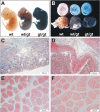
Analysis of the expression of Hif-p4h-2 in embryos and adult tissues. Hif-p4h-2 promoter-driven β-galactosidase expression was analyzed by X-gal staining of whole mounts or 5-μm paraffin sections of wild-type (wt), Hif-p4h-2+/gt (wt/gt), and Hif-p4h-2gt/gt (gt/gt) embryos and different tissues from adult mice. Embryonic day 12.5 Hif-p4h-2+/gt and Hif-p4h-2gt/gt embryos (A) and whole mount hearts (B) show ubiquitous X-gal staining with genotype-dependent intensity. C–F, histological analysis demonstrates pronounced X-gal staining in the cardiomyocytes of the Hif-p4h-2gt/gt hearts (C), in the smooth muscle cells of Hif-p4h-2gt/gt pulmonary arterial walls (D), and in myocytes of skeletal muscle (E), whereas no staining was observed in wild-type skeletal muscle (F).
FIGURE 3.
FIGURE 3.
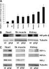
Analysis of wild-type Hif-p4h-2 mRNA levels and Hif-p4h-2, Hif-1α, and Hif-2α protein levels in selected tissues. A, wild-type Hif-p4h-2 mRNA levels in various Hif-p4h-2gt/gt tissues relative to wild-type tissues were analyzed by Q-PCR with primers amplifying the wild-type cDNA. A significant decrease (p < 0.001, n ≥ 5) in the Hif-p4h-2 mRNA level was seen in all Hif-p4h-2gt/gt tissues studied relative to the wild type, except in the liver. The error bars indicate S.D. B and C, Western blotting of total protein extracts from wild-type (wt) and Hif-p4h-2gt/gt (gt/gt) heart, skeletal (Sk) muscle, and kidney was performed with the antibodies indicated. Tubulin and HIF-1β stainings were used as loading controls.
FIGURE 4.
FIGURE 4.
Analysis of hematological parameters, serum Epo, and Epo mRNA in the kidney. Hematocrit, the red blood cell count, and hemoglobin level were analyzed in blood samples from four adult Hif-p4h-2gt/gt and age- and gender-matched wild-type mice. Serum Epo levels in six adult Hif-p4h-2gt/gt and age- and gender-matched wild-type mice were analyzed by enzyme-linked immunosorbent assay. The relative amounts of Epo mRNA in kidney samples from eight to ten adult Hif-p4h-2gt/gt and age- and gender-matched wild-type mice were analyzed by Q-PCR. The error bars indicate S.D.
FIGURE 5.
FIGURE 5.
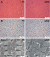
Histological analyses of the hearts. A and B, 5-μm paraffin sections of wild-type (wt) and Hif-p4h-2gt/gt (gt/gt) hearts stained with hematoxylin and eosin (A) and Pecam-1 (B). C, transmission electron microscopy of the wild-type (wt) and Hif-p4h-2gt/gt (gt/gt) hearts. The intensity and distance between Z-bands are influenced by the contractile state and sample width and vary within the same samples irrespective of the genotype.
FIGURE 6.
FIGURE 6.
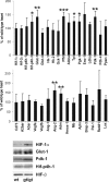
Molecular analysis of the preischemic Hif-p4h-2gt/gt hearts. Top and middle panels, Q-PCR analysis of the mRNA levels of Hif-1α, Hif-2α, Hif-p4h-1 and 3, and certain HIF target and other genes in adult preischemic Hif-p4h-2gt/gt hearts relative to age- and gender-matched wild-type hearts (n = 6). Hk-1 and -2, hexokinase-1 and -2; Gck-1, glucokinase-1; Pfkl, phosphofructokinase-L; Aldoa, aldolase A; Tpi, triose phosphate isomerase; Pgk-1, phosphoglycerate kinase-1; Eno-1, enolase-1; Ldha, lactate dehydrogenase A; A2bar, adenosine receptor A2b; A3ar, adenosine receptor A3; Adm, adrenomedullin; Hmox1, heme oxygenase 1; Mb, myoglobin; Apln, apelin; Pai-1, plasminogen activator inhibitor-1; Lox, lysyl oxidase. *, p < 0.05; **, p < 0.01; ***, p < 0.001. The error bars indicate S.D. Bottom panels, Western blotting of total protein extracts from preischemic wild-type (wt) and Hif-p4h-2gt/gt (gt/gt) heart was performed with the antibodies indicated.
FIGURE 7.
FIGURE 7.
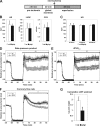
Analysis of Hif-p4h-2gt/gt hearts in an ex vivo ischemia-reperfusion model. A, hearts from 2–5-month-old female Hif-p4h-2gt/gt (gt/gt) mice and age- and gender-matched controls (wt) were isolated and subjected to Langendorff perfusion as indicated. B, preischemic heart rate (HR), LVDP, and coronary flow rate (CFR) were measured after a minimum 30-min stabilization period. C, post-ischemic heart rates during 0–15, 15–30, and 30–45 min of reperfusion. D–F, global ischemia (black horizontal bar) was induced after the stabilization perfusion (the last 10 min of which is shown) by cessation of perfusion for 20 min. Ischemia was followed by 45 min of reperfusion. The values are the means, and the hatched area depicts ± S.E. Coronary flow rate and left ventricle pressure were continuously recorded, and HR, LVDP, and RPP were calculated. The post-ischemic RPP (D), dP/dtmax (E), and coronary flow rate (F) values were calculated relative to the preischemic mean (mean of RPP, dP/dtmax, and coronary flow rate for the last 3 min of stabilization) in the wild-type (n = 7) and Hif-p4h-2gt/gt (n = 9) hearts. The AUC during the 45-min post-ischemic period was calculated using the method of summary measures. *, p < 0.05; **, p < 0.01. G, lactate dehydrogenase (LDH) levels in the venous effluents collected during the reperfusion were determined, and the cumulative lactate dehydrogenase washout for the first 15 min of reperfusion was calculated (units/g of wet weight) for the wild-type (n = 8) and Hif-p4h-2gt/gt (n = 8) hearts. The error bars indicate S.E.
FIGURE 8.
FIGURE 8.
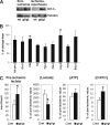
Molecular and metabolic analysis of Hif-p4h-2gt/gt hearts. A, Western blotting of total protein extracts from wild-type (wt) and Hif-p4h-2gt/gt (gt/gt) heart for HIF-1α at end ischemia and after ischemia-reperfusion. Tubulin staining was used as a loading control. B, Q-PCR analysis of the mRNA levels of certain HIF target genes (see Fig. 6) in Hif-p4h-2gt/gt hearts relative to age- and gender-matched wild-type hearts after ischemia-reperfusion (n = 4). The error bars indicate S.D. C, the amount of preischemic lactate (n = 4), and the ratios of the end ischemic versus preischemic [Lactate], [ATP], and [CrP/Cr] (n = 3) in age- and gender-matched Hif-p4h-2gt/gt and wild-type hearts. The error bars indicate S.D.

Similar articles

Cited by

References

    1. Kaelin W. G., Jr., Ratcliffe P. J. (2008) Mol. Cell 30, 393–402 - PubMed
    1. Fraisl P., Aragonés J., Carmeliet P. (2009) Nat. Rev. Drug Discov. 8, 139–152 - PubMed
    1. Semenza G. L. (2009) Physiology 24, 97–106 - PubMed
    1. Ivan M., Kondo K., Yang H., Kim W., Valiando J., Ohh M., Salic A., Asara J. M., Lane W. S., Kaelin W. G., Jr. (2001) Science 292, 464–468 - PubMed
    1. Jaakkola P., Mole D. R., Tian Y. M., Wilson M. I., Gielbert J., Gaskell S. J., Kriegsheim Av, Hebestreit H. F., Mukherji M., Schofield C. J., Maxwell P. H., Pugh C. W., Ratcliffe P. J. (2001) Science 292, 468–472 - PubMed

Publication types

MeSH terms

Substances