

doi: 10.1167/iovs.09-4396.
Synaptic organization of the vertebrate retina: general principles and species-specific variations: the Friedenwald lecture
Affiliations
- PMID: 20185835
- PMCID: PMC3258978
- DOI: 10.1167/iovs.09-4396
Item in Clipboard
Synaptic organization of the vertebrate retina: general principles and species-specific variations: the Friedenwald lecture
Invest Ophthalmol Vis Sci.
2010 Mar.
No abstract available
Figures
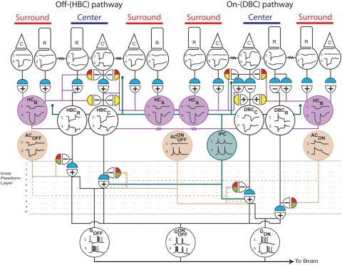
Summary of the major synapses that mediate the CSARF of BCs and GCs in the retina. Left: the OFF- or HBC-pathway; right: the ON- or DBC-pathway. Top traces: the voltage response to center illumination; bottom traces: the response to surround illumination. In the postsynaptic semicircles, +, sign-preserving chemical synapses; −, sign-inverting chemical synapses; zigzags, electric synapses. Neurotransmitter color code in the presynaptic semicircles: blue: glutamatergic; red: GABAergic; green: glycinergic; and yellow: unspecified. R, rod; C, cone; HC, horizontal cell (A-type: HCA and B-type: HCB); HBCR, HBCC, DBCR, and DBCC are rod- and cone-dominated hyperpolarizing and depolarizing BCs; AON, sustained ON amacrine cell; AON-OFF, transient ON-OFF amacrine cell; AOFF, sustained OFF amacrine cell; IPC, interplexiform cell; GON, sustained ON ganglion cell; GON-OFF, ON-OFF ganglion cell; GOFF, sustained OFF ganglion cell; OPL, outer plexiform layer; IPL, inner plexiform layer. For animation of the center-surround signaling pathways, please visit http://neuro.neusc.bcm.tmc.edu/wu/resources.html .
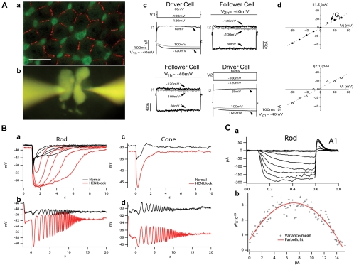
Electrical coupling and HCN1 channels in salamander photoreceptors. (Aa) Confocal image of a salamander flatmount retina (with the focal plane at the level of the distal region of rod cell bodies) double labeled with anti-Cx35/36 (red) and recoverin (green). Recoverin differentially labeled rods (r, weak green) and cone inner segments (c, strong green). The strong Cx35/36 punctate labeling on membrane contacts outlined the mosaic of the rod network in the field. Scale bar, 20 μm. (Ab) A pair of rods simultaneously patch clamped and filled with Lucifer yellow through two recording pipettes. (Ac) Simultaneous dual whole-cell voltage clamp recordings from a pair of neighboring rods. The membrane potential of two rods was held at −40 mV. Upper panel: when a series of voltage step commands (V1) (from −120 mV to 60 mV with an increment of 20 mV) were applied to cell 1 (driver cell), the voltage activated current responses (I1) (arrowheads) were recorded in cell 1 (left panel) and the junctional currents of the opposite polarity (I2) (arrows) were recorded in cell 2 (follower cell, right panel). Lower panel: switching the position of driver/follower cells. (Ad) Relations of transjunctional current (Ij) and transjunctional voltage (Vj) obtained in upper and lower panels in (Ac). The junctional conductance (Gj) measured in either direction is 500 pS. (Reprinted from Zhang J, Wu SM. Physiological properties of rod photoreceptor electrical coupling in the tiger salamander retina. J Physiol. 2005;564:849–86. © 2005 by The Physiological Society.) (Ba) Rod responses to flashes of increasing light intensity in normal Ringer's (black traces) and in the presence of 100 μM HCN channel blocker ZD 7288 (red traces). (Bb) Rod response to a frequency-chirped light stimulus (chirped sine wave-modulated light ranged from 0.5 to 5 Hz over the course of 20 seconds) in normal Ringer's (black) and in 100 μM ZD 7288 (red). (Bc, Bd) Cone responses to flashes of increasing light intensity (Bc) and to frequency-chirped light stimulus (Bd) in normal Ringer's (black traces) and in the presence of 100 μM ZD 7288 (red traces). (Ca) Whole-cell recording of HCN channels from a rod to hyperpolarizing voltage steps with an extracellular solution containing TEA, cobalt and barium so that all other ionic currents other than Ih were blocked. (Cb) Variance versus mean plot computed from an ensemble of whole-cell Ih current. The slope of the variance-mean plot at 0 mean current gives an estimate of single channel conductance of 663 ± 71 fS, and the peak of the hyperbolic curve gives an estimate of the total number of HCN1 channels per rod of 2214 ± 986. (Part C reprinted with permission from Barrow AJ, Wu SM. Low-conductance HCN1 ion channels augment the frequency response of rod and cone photoreceptors. J Neurosci. 2009;29:5841–5853. © 2009 by the Society for Neuroscience.)
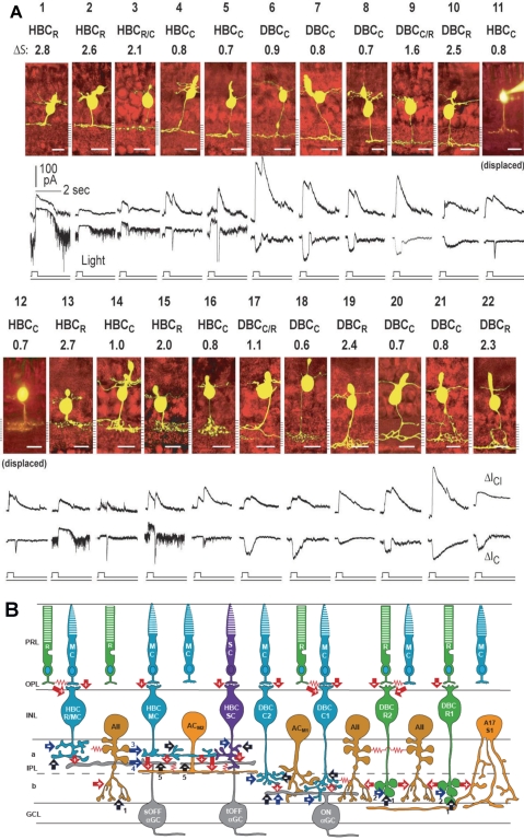
(A) Stratum-by-stratum rules for correlating patterns of axon terminal ramification and physiological responses in retinal bipolar cells. 22 morphologically identified (by Lucifer yellow filling) BCs and their light-evoked excitatory cation current (ΔIC) and inhibitory chloride current (ΔICl) recorded from dark-adapted salamander retinal slices. Each cell is named according to their spectral difference (ΔS) and ΔIC polarity as rod-dominated, cone-dominated, mixed rod/cone hyperpolarizing or depolarizing bipolar cells (HBCR, HBCC, HBCR/C, DBCR, DBCC or DBCR/C). BCs with inward ΔIC are DBCs and with outward ΔIC are HBCs. The spectral difference, ΔS, is defined as S700 − S500 (where S700 and S500 are intensities in log units of 700- and 500-nm light eliciting responses of the same amplitude). Since ΔS for the salamander rods is approximately 3.4 and that for the cones is approximately 0.1, BCs with ΔS > 2.0 are rod-dominated (HBCR or DBCR), with ΔS < 1.0 are cone-dominated (HBCC or DBCC), and with 1.0 < ΔS < 2.0 are mixed rod/cone cells (HBCR/C or DBCR/C, also named HBCM or DBCM, see Fig. 4). Displaced HBCCs: HBCCs with somas displaced in the outer nuclear layer. (Modified from Pang J-J, Gao F, Wu SM. Stratum-by-stratum projection of light response attributes by retinal bipolar cells of Ambystoma. J. Physiol. 2004;558:249–262. © 2004 by The Physiological Society; and Maple BR, Zhang J, Pang J-J, Gao F, Wu SM. Characterization of displaced bipolar cells in the tiger salamander retina. Vision Res. 2005;45:697–705. © 2005 Elsevier Ltd.) (B) Schematic diagram of synaptic connections of photoreceptors, BCs, ACs, and α GCs in the mammalian retina. R, rod; MC, M-cone; SC, S-cone; HBCMC/R, mixed M-cone/rod hyperpolarizing BC; HBCMC, M-cone dominated hyperpolarizing bipolar cell; HBCSC, S-cone dominated hyperpolarizing bipolar cell; DBCC2, type 2 cone depolarizing bipolar cell; DBCC1, type 1 cone depolarizing bipolar cell; DBCR2, type 2 rod depolarizing bipolar cell; DBCR1, type 1 rod depolarizing bipolar cell. Note that BCs with the most rod inputs have axon terminal endings near the two margins of the IPL, whereas those with the most cone inputs bear axons ramifying in the central regions of the IPL, similar to the rules set forward by the salamander BCs (A). ACM1, M-cone dominated depolarizing amacrine cell; ACM2, M-cone dominated ON-OFF amacrine cell; AII, AII amacrine cell; A17/S1, A17 amacrine cell; sOFFαGC, sustained OFF αGC; tOFFαGC, transient OFF αGC; ONαGC, ON αGC; green: rods and rod BCs; blue: M-cones and M-cone BCs, purple: S-cone and S-cone BCs; light orange: GABAergic ACs; dark orange: glycinergic ACs; gray: αGCs; arrows: chemical synapses (red, glutamatergic; black, GABAergic; blue, glycinergic; +, sign-preserving; −, sign-inverting); zigzag (red): electrical synapses. PRL, photoreceptor layer; OPL, outer plexiform layer; INL, inner nuclear layer; IPL, inner plexiform layer (a, sublamina a, b, sublamina b); GCL, ganglion cell layer. Many inhibitory synapses from unspecified ACs are represented as black and blue arrows.
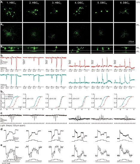
Morphology, light responses, and receptive fields of six types of bipolar cells in the tiger salamander retina. (a) Fluorescent micrographs of a neurobiotin-filled HBCR (column 1), a HBCM (column 2), HBCC (column 3), a DBCC (column 4), a DBCM (column 5), and a DBCR (column 6) viewed with a confocal microscope at the outer INL/OPL level (ai), the IPL level (aii), and with z-axis rotation (aiii). Calibration bar, 100 μm. (bi) BC voltage responses to 500-nm and 700-nm light steps of various intensities. (bii) Response-intensity (V-Log I) curves of the responses to 500-nm and 700-nm lights. ΔS (spectral difference, see Fig. 3A) of the 6 BCs are 2.13, 1.51, 0.30, 0.57, 1.45, and 2.25. (c) Measurements of BC receptive field center diameters (RFCD) by recording voltage responses to a 100-μm-wide light bar moving stepwise (with 120-μm step increments) across the receptive field. (d) Voltage responses of the 6 types of BCs elicited by a center light spot (300 μm) and a surround light annulus (700 μm inner diameter, 2000 μm outer diameter). The surround light annulus was of the same intensity (700 nm, −2) for all 6 cells whereas the intensity of the center light spot was adjusted so that it allowed the annulus to produce the maximum response. (e) Voltage responses of the 6 types of BCs elicited by a center light spot and a surround light annulus (same as in d), and by a train of −0.1-nA/200-msec current pulses passed into the cell by the recording microelectrode through a bridge circuit. (Reprinted with permission from Zhang A-J, Wu SM. Receptive fields of retinal bipolar cells are mediated by heterogeneous synaptic circuitry. J Neurosci. 2009;29:789–797. © 2009 by The Society for Neuroscience.)
Similar articles
-
Photoreceptor membrane proteins, phototransduction, and retinal degenerative diseases. The Friedenwald Lecture.Invest Ophthalmol Vis Sci. 1998 Dec;39(13):2491-513. Invest Ophthalmol Vis Sci. 1998. PMID: 9856758 Review. No abstract available.
-
Photopigments and seeing--lessons from natural experiments: the Proctor lecture.Invest Ophthalmol Vis Sci. 1998 Nov;39(12):2204-16. Invest Ophthalmol Vis Sci. 1998. PMID: 9804128 Review. No abstract available.
-
Keeping an eye on the time: the Cogan Lecture.Invest Ophthalmol Vis Sci. 2002 May;43(5):1286-98. Invest Ophthalmol Vis Sci. 2002. PMID: 11980836 No abstract available.
-
G protein signaling in the retina and beyond: the Cogan lecture.Invest Ophthalmol Vis Sci. 2014 Dec 15;55(12):8201-7. doi: 10.1167/iovs.14-15928. Invest Ophthalmol Vis Sci. 2014. PMID: 25511392 Free PMC article. No abstract available.
-
Rhodopsin structure, function, and topography the Friedenwald lecture.Invest Ophthalmol Vis Sci. 2001 Jan;42(1):3-9. Invest Ophthalmol Vis Sci. 2001. PMID: 11133841 Review. No abstract available.
Cited by
-
Quantitative biology of single neurons.J R Soc Interface. 2012 Dec 7;9(77):3165-83. doi: 10.1098/rsif.2012.0417. Epub 2012 Aug 22. J R Soc Interface. 2012. PMID: 22915636 Free PMC article. Review.
-
Assessment of area and structural irregularity of retinal layers in diabetic retinopathy using machine learning and image processing techniques.Sci Rep. 2024 Feb 18;14(1):4013. doi: 10.1038/s41598-024-54535-6. Sci Rep. 2024. PMID: 38369610 Free PMC article.
-
Characterization and staging of outer plexiform layer development in human retina and retinal organoids.Development. 2021 Dec 1;148(23):dev199551. doi: 10.1242/dev.199551. Epub 2021 Dec 8. Development. 2021. PMID: 34738615 Free PMC article.
-
Olfactomedin-like 2 A and B (OLFML2A and OLFML2B) profile expression in the retina of spotted gar (Lepisosteus oculatus) and bioinformatics mining.Fish Physiol Biochem. 2019 Oct;45(5):1575-1587. doi: 10.1007/s10695-019-00647-0. Epub 2019 May 20. Fish Physiol Biochem. 2019. PMID: 31111317
-
Human Occipital and Parietal GABA Selectively Influence Visual Perception of Orientation and Size.J Neurosci. 2017 Sep 13;37(37):8929-8937. doi: 10.1523/JNEUROSCI.3945-16.2017. Epub 2017 Aug 14. J Neurosci. 2017. PMID: 28821653 Free PMC article.
References
-
- Cajal SR. La retine des vertebres. La Cellule. 1893;9:17–257
-
- Baylor DA. Photoreceptor signals and vision: The Proctor Lecture. Invest Ophthalmol Vis Sci. 1987;28:34–49 - PubMed
-
- Yau KW. Phototransduction mechanism in retinal rods and cones: The Friedenwald Lecture. Invest Ophthalmol Vis Sci. 1994;35:9–32 - PubMed
-
- Dowling JE. The Retina, an Approachable Part of the Brain. Boston: Harvard University Press; 1987.
-
- Wassle H. Parallel processing in the mammalian retina. Nat Rev Neurosci. 2004;5:747–757 - PubMed
Publication types
MeSH terms
Grants and funding
LinkOut - more resources
Full Text Sources
