. 2010 Mar 17;29(6):1116-25.
doi: 10.1038/emboj.2010.14. Epub 2010 Feb 25.

A functional peptidyl-tRNA hydrolase, ICT1, has been recruited into the human mitochondrial ribosome

Affiliations

A functional peptidyl-tRNA hydrolase, ICT1, has been recruited into the human mitochondrial ribosome

Ricarda Richter et al. EMBO J. .

Abstract

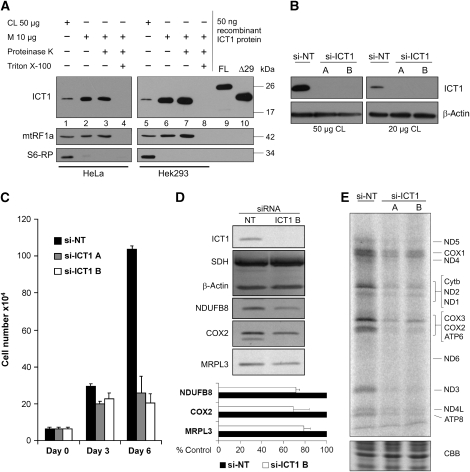
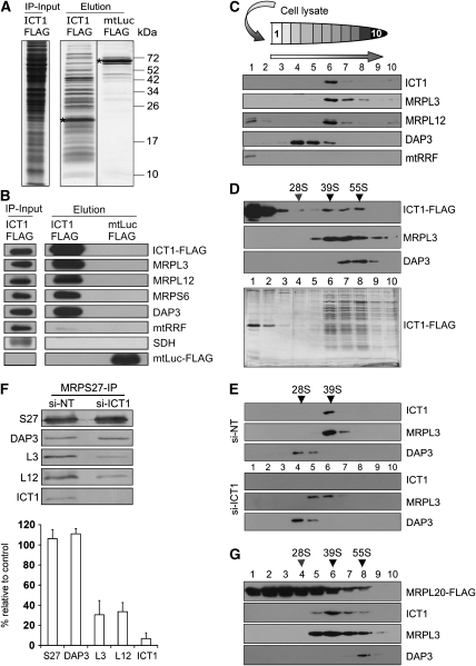
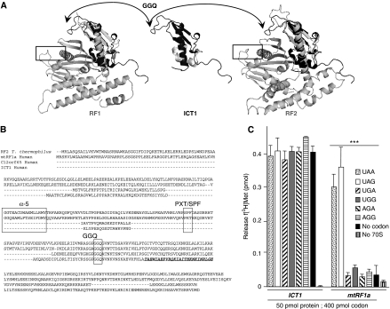
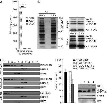
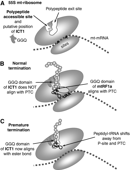
Bioinformatic analysis classifies the human protein encoded by immature colon carcinoma transcript-1 (ICT1) as one of a family of four putative mitochondrial translation release factors. However, this has not been supported by any experimental evidence. As only a single member of this family, mtRF1a, is required to terminate the synthesis of all 13 mitochondrially encoded polypeptides, the true physiological function of ICT1 was unclear. Here, we report that ICT1 is an essential mitochondrial protein, but unlike the other family members that are matrix-soluble, ICT1 has become an integral component of the human mitoribosome. Release-factor assays show that although ICT1 has retained its ribosome-dependent PTH activity, this is codon-independent; consistent with its loss of both domains that promote codon recognition in class-I release factors. Mutation of the GGQ domain common to ribosome-dependent PTHs causes a loss of activity in vitro and, crucially, a loss of cell viability, in vivo. We suggest that ICT1 may be essential for hydrolysis of prematurely terminated peptidyl-tRNA moieties in stalled mitoribosomes.


Conflict of interest statement

The authors declare that they have no conflict of interest.

Figures

Figure 1
Figure 1
ICT1 is an essential protein necessary for mitochondrial protein synthesis. (A) Human ICT1 is a mitochondrial protein. Cell lysate (CL 50 μg, lanes 1, 5) or mitochondria (10 μg, lanes 2–4, 6–8) were isolated from HeLa and HEK293T cells and subjected to western blot analysis either immediately (lanes 1, 2; 5, 6) or after treatment with proteinase-K (lanes 3, 7). Mitochondria were lysed with Triton X-100 to confirm the sensitivity of marker proteins to the protease (lanes 4, 8). Mitochondrial release factor-1a (mtRF1a) was used as a mitochondrial matrix marker and ribosomal protein-S6 (S6-RP) as a cytosolic marker. Purified FL (lane 9) and an ICT1 deleted of N-terminal 29 residues (Δ29, lane 10) are shown in comparison with the endogenous protein. (BE) Depletion of ICT1 inhibits cell growth, impairs mitochondrial protein synthesis and decreases mitochondrial respiratory chain complexes. HeLa cells in standard glucose media were transfected with either of two siRNAs directed to ICT1 transcript (si-ICT1A or B) or a non-targeting control (si-NT), and cell numbers were counted at 3-day intervals. Standard errors were derived from three independent experiments (C). Cell lysates were isolated from non-targeting and ICT1-depleted cells (3 days) and subjected to western blotting for ICT1 (B) and various markers (D). The relative levels for the MRP MRPL3 and respiratory components NDUFB8 or COX2 were quantified (lower panel) with standard errors derived from three independent repeats. (E) After 3-day siRNA-mediated depletion, cells were subjected to metabolic labelling of mitochondrial proteins for 15 min after inhibition of cytosolic protein synthesis. Aliquots (50 μg) were separated by 15% SDS–PAGE and exposed to a PhosphorImager. Proteins are identified by comparison against those reported by Chomyn (1996). A section of the gel stained with Coomassie blue (CBB) following exposure is shown to indicate even loading of cell lysate.
Figure 2
Figure 2
ICT1 is an integral component of the mitoribosome. (A, B) FLAG-tagged ICT1 immunoprecipitates mitoribosomes. HEK293T cells expressing FLAG-tagged ICT1 or mitochondrially localised luciferase (mtLuc-FLAG) were induced for 3 days; mitochondria were isolated, lysed and subjected to IP as detailed. The eluate and mitochondrial lysate before IP (IP-input) were separated by 15% SDS–PAGE and visualised by silver staining. * designates the FLAG protein. (B) Aliquots of the eluates were also subjected to western blot analysis with the indicated antibodies: MRPL3, MRPL12, MRPS6 and DAP3 as mitoribosomal markers; mtRRF, mitoribosome recycling factor; SDH, 70-kDa component of complex-II. (C) ICT1 co-sediments with the large mitoribosomal subunit. HeLa cells were lysed (600 μg), separated through a 10–30% sucrose gradient and fractionated as detailed (HeLa and HEK293T lysates gave identical separations). Components of the 39S mt-LSU (MRPL3, MRPL12) and 28S mt-SSU (DAP3) mitoribosomal subunits were visualised by western blotting. On immediate lysis, mtRRF is used as a matrix-soluble marker. (D) ICT1 also co-sediments with the intact monosome. Mitochondria (3 mg) of ICT1-FLAG-expressing HEK293T cells were subjected to FLAG IP; the entire eluate was separated by isokinetic density gradients and fractions were blotted as detailed above or visualised by silver staining (lower panel). Mitochondrial SSU (DAP3) and mt-LSU (MRPL3) MRPs are visualised. The approximate indicators for 28S mt-SSU, 39S mt-LSU and 55S monosome are shown and were determined as described under Materials and methods. (E) ICT1 is an integral member of 39S mt-LSU. Cell lysates (600 μg) from ICT1-depleted (si-ICT1B) or non-targeted control cells (si-NT) were separated by isokinetic gradients and proteins were visualised in the fractions by western blotting as described. Sedimentation markers were identified as above. (F) Loss of ICT1 causes depletion of the monosome. Cells expressing MRPS27-FLAG were treated with si-NT or si-ICT1B, after which IP was performed. To assess monosome formation, levels of MRPL3 and MRPL12 were quantified by western blotting of three individual experiments (right panel; MRPL3 P=0.001, MRPL12 P<0.001, MRPS27 P=0.3). (G) ICT1's association with mitoribosomes is not FLAG-dependent. Mitochondria from cells expressing MRPL20-FLAG were subjected to FLAG IP and the eluate was analysed by western blotting after isokinetic density gradients as described in panel D.
Figure 3
Figure 3
ICT1 is a codon-independent PTH. (A) Structural comparisons between ICT1 and members of RF1 or RF2. Only limited structure is available for murine ICT1 (centre, PDB 1J26 unpublished structural genomics output), but this can be superimposed using Topmatch (Sippl and Wiederstein, 2008) onto either RF family (left T. maritima PDB 1RQ0 (Shin et al, 2004); right T. thermophilus PDB 2IHR (Zoldak et al, 2007)), making it unclear as to its evolutionary origin. The arrows show the ICT 1GGQ motif and the boxes enclose the codon recognition domains (Ito et al, 2000; Laurberg et al, 2008). (B) Primary sequence comparisons of ICT1 and translation release factor members. Representatives of the release factor families are shown; RF1 (human mtRF1a), RF2 (T. thermophilus) aligned with human sequences for ICT1 and a fourth member of the mitochondrial release factor family, C12orf65. Three regions are highlighted; the GGQ motif conferring PTH activity, and α-5/tripeptide domains that are implicated in codon recognition. The latter two domains are absent in ICT1 and C12orf65. There are two other families of PTHs, PTH1 and PTH2 (reviewed by Das and Varshney, 2006), which are predicted to be represented in the human mitochondrion by PTRH1 and PTRH2 (uniprot Q86Y79 and Q9Y3E5, respectively). These protein families function independently of the ribosome and on the basis of comparison of their three-dimensional structures (data not shown), are not homologous to the ribosome-dependent PTH family that contains ICT1. (C) ICT1 has codon-independent and ribosome-dependent translation release factor activity. E. coli ribosomes were programmed with tritiated P-site fmet-tRNAMet and A-site codons as indicated (detailed under Materials and methods). Activity was measured as hydrolysis of f[3H]met from its cognate tRNAMet and is represented as pmol f[3H]met released. Non-limiting amounts of protein (50 pmol) and RNA triplet (400 pmol) were used in the assay where required, with mtRF1a as a positive control. Activities are also evident where ribosomes were programmed with no codon or were absent from the assay, entirely. Reactions lacking 70S ribosomes contained the UAA triplet. Standard errors were calculated from a minimum of eight repeats; ***P<0.001.
Figure 4
Figure 4
Mutations of the GGQ domain can affect cell viability. (A) GGQ-mutant derivatives of ICT1 have lost PTH activity. Wild type and mutant derivatives (AGQ, GSQ) of Δ29 ICT1 were expressed as GST-fusion proteins, cleaved and assayed for f[3H]met release as described in Figure 3. All assays were performed with UAG codons and purified proteins, all equally monodispersed as assessed by dynamic light scattering (data not shown); ***P<0.001. (B, C) Mutated ICT1 is assembled into the mitoribosome. (B) FLAG-tagged wild-type (GGQ) and mutant (GSQ) ICT1 were expressed in HEK293T cells and the eluate from FLAG IP was subjected to silver staining (left panel) or western blotting (right panels) after denaturing gel electrophoresis. Molecular weight markers are indicated. The western blots of mitochondrial lysates shown are those before (IP-input) and after (Elution) FLAG IP of the wild type and mutated ICT1 derivatives. (C) Cell lysates were subjected to isokinetic gradient analysis before fractionation and western blotting, as described. The upper three panels are from wild-type ICT1-FLAG, the middle panels from mutated GSQ ICT1-FLAG and the lower panels from control mtLuc-FLAG. (D) A mutation of the GGQ domain affects cell growth. Non-targeting si-RNA-treated cells served as negative control (1, WT si-NT). Cells with only endogenous ICT1 (WT), or overexpressing normal (GGQ) or mutated (GSQ) ICT1 were treated for 4 days with 10 nM si-ICT1B to deplete endogenous ICT1 (2–4), whereas lane-2 represented the fully depleted control (WT si-ICT1B). Growth rates were compared by counting populations after 3 days of siRNA treatment; 3 versus 4: P<0.01; 1 versus 4: P<0.001. Western blots of lysates (4 days of siRNA treatment) after interrogation with the indicated antibodies are shown to the right.
Figure 5
Figure 5
A schematic representation of the putative position and function of ICT1 in the human mitochondrial ribosome. (A) A simplified cartoon of the 55S mitochondrial ribosome indicating the polypeptide exit tunnel and site (PES) and the PAS in the large subunit as defined by Sharma et al (2003). No orthologues of the proteins that would occupy the PAS have been found in mammalian mitochondria, and we postulate that is where ICT1 is positioned with the GGQ domain inserted deep into the pocket. Sites for the aminoacyl (A) and peptidyl (P) tRNAs are shown. The mt-mRNA is depicted between the large and small mt-ribosomal subunits. (B) Under conditions of normal termination, the ester bond of the peptidyl-tRNA is positioned close to the peptidyl-transferase centre (PTC); the release factor, mtRF1a, enters through the A-site, recognising the stop codon (UAA) and aligning the GGQ domain at the PTC to promote hydrolysis of the ester bond and release of the nascent peptide. (C) Where abortive elongation occurs, the peptidyl-tRNA may drop away from the P-site towards the PES, aligning the ester bond close to the GGQ domain of ICT1, promoting cleavage of the tRNA, which allows both mt-tRNA and truncated peptide to be released from the mitochondrial monosome (or potentially from dissociated 39S mt-LSU).

Comment in

Similar articles

Cited by

References

    1. Caskey CT, Beaudet AL, Scolnick EM, Rosman M (1971) Hydrolysis of fMet-tRNA by peptidyl transferase. Proc Natl Acad Sci USA 68: 3163–3167 - PMC - PubMed
    1. Chomyn A (1996) In vivo labeling and analysis of human mitochondrial translation products. Methods Enzymol 264: 197–211 - PubMed
    1. Chrzanowska-Lightowlers ZM, Preiss T, Lightowlers RN (1994) Inhibition of mitochondrial protein synthesis promotes increased stability of nuclear-encoded respiratory gene transcripts. J Biol Chem 269: 27322–27328 - PubMed
    1. Das G, Varshney U (2006) Peptidyl-tRNA hydrolase and its critical role in protein biosynthesis. Microbiology 152: 2191–2195 - PubMed
    1. Frolova LY, Tsivkovskii RY, Sivolobova GF, Oparina NY, Serpinsky OI, Blinov VM, Tatkov SI, Kisselev LL (1999) Mutations in the highly conserved GGQ motif of class 1 polypeptide release factors abolish ability of human eRF1 to trigger peptidyl-tRNA hydrolysis. RNA 5: 1014–1020 - PMC - PubMed

Publication types

MeSH terms