. 2010 Jul;17(7):492-500.
doi: 10.1038/cgt.2010.3. Epub 2010 Feb 26.

Enhanced anti-tumor effects of combined MDR1 RNA interference and human sodium/iodide symporter (NIS) radioiodine gene therapy using an adenoviral system in a colon cancer model

Affiliations

Enhanced anti-tumor effects of combined MDR1 RNA interference and human sodium/iodide symporter (NIS) radioiodine gene therapy using an adenoviral system in a colon cancer model

S J Ahn et al. Cancer Gene Ther. 2010 Jul.

Abstract

Using an adenoviral system as a delivery mediator of therapeutic gene, we investigated the therapeutic effects of the use of combined MDR1 shRNA and human NIS (hNIS) radioiodine gene therapy in a mouse colon xenograft model. In vitro uptake of Tc-99m sestamibi was increased approximately two-fold in cells infected with an adenovirus vector that expressed MDR1 shRNA (Ad-shMDR1) and I-125 uptake was 25-fold higher in cells infected with an adenovirus vector that expressed human NIS (Ad-hNIS) as compared with control cells. As compared with doxorubicin or I-131 treatment alone, the combination of doxorubicin and I-131 resulted in enhanced cytotoxicity for both Ad-shMDR1- and Ad-hNIS-infected cells, but not for control cells. In vivo uptake of Tc-99m sestamibi and Tc-99m pertechnetate was twofold and 10-fold higher for Ad-shMDR1 and Ad-hNIS-infected tumors as compared with tumors infected with a control adenovirus construct that expressed beta-galactosidase (Ad-LacZ), respectively. In mice treated with either doxorubicin or I-131 alone, there was a slight delay in tumor growth as compared to mice treated with Ad-LacZ. However, combination therapy with doxorubicin and I-131 induced further significant inhibition of tumor growth as compared with mice treated with Ad-LacZ. We have shown successful therapeutic efficacy of combined MDR shRNA and hNIS radioiodine gene therapy using an adenoviral vector system in a mouse colon cancer model. Adenovirus-mediated cancer gene therapy using MDR1 shRNA and hNIS would be a useful tool for the treatment of cancer cells expressing multi-drug resistant genes.


Figures
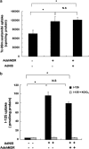
Figure 1
Figure 1
Functional uptake of Tc-99m sestamibi and I-125 in Ad-shMDR1 or Ad-hNIS-infected HCT-15 cells. HCT-15 cells were infected with Ad-shMDR1 (200 MOI) or Ad-shMDR1 (200 MOI) plus Ad-hNIS (20 MOI) for 48 h and were incubated with 370 kBq Tc-99m sestamibi for 30 min (a). I-125 uptake was performed in transfected HCT15 cells with Ad-hNIS (20 MOI) or Ad-hNIS (20 MOI) plus Ad-shMDR1 (100 MOI). I-125 uptake was then initiated for 30 min by the addition of 0.5 ml HBSS containing 3.7 kBq of I-125 per well and was blocked in the presence of potassium perchlorate, a known inhibitor of NIS function (b). Results are expressed as the mean of c.p.m. per protein concentration. The data shown are the means of experiments performed in triplicate. Bars represent means±s.d.; NS, not significant; *statistically significant.
Figure 2
Figure 2
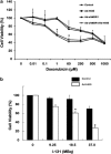
Cytotoxic effects of doxorubicin or I-131 in Ad-shMDR1 and Ad-hNIS-infected cancer cells. HCT-15 cells (infected with HBSS, 20 MOI Ad-hNIS, 200 MOI Ad-shMDR1 and both 200 MOI Ad-shMDR1 and 20 MOI Ad-hNIS) were incubated in the presence of 0, 0.01, 0.1, 1, 60, 250, 500 and 1000 μM doxorubicin for 48 h at 37 °C (a). HC-T15 cells infected with 20 MOI of Ad-hNIS were treated with 0, 9.25, 18.5 and 37.0 MBq of I-131 in HBSS or HBSS alone for 7 h. Cellular viability assessed by the use of a clonogenic assay was compared to treatment with I-131 or with HBSS alone (b). The data shown are the means of experiments performed in triplicate. Bars represent means±s.d.; NS, not significant; *statistically significant
Figure 3
Figure 3
Cellular cytotoxic effects of combined doxorubicin and I-131 in both Ad-shMDR1 and Ad-hNIS-infected HCT15 cells. HCT-15 cells were infected with both Ad-shMDR1 (200 MOI) and Ad-hNIS (20 MOI) for 48 h. At 48 h after viral infection, cells were treated with HBSS, 9.25 and 18.5 MBq of I-131 for 7 h. I-131-treated cells (1000 cells per well) were incubated with or without 1 or 10 μM doxorubicin at 37 °C for 1 week. Cells were stained with a crystal violet solution and colonies of more than 30 cells were counted. Results are expressed as the percentage of surviving colonies. The data shown are the means of experiments performed in triplicate. Bars represent means±s.d.
Figure 4
Figure 4
In vivo imaging of tumor-bearing mice infected with both Ad-shMDR1 and Ad-hNIS. A total of 1 × 10⁷ HCT-15 cells were s.c injected into both thighs of mice. When the tumor volume reached approximately 100 mm3, Ad-shMDR1 (2 × 10⁹ PFU) and Ad-hNIS (1 × 10⁹ PFU) were intratumorally injected into the tumor-bearing mice. Ad-LacZ as a control was intratumorally injected to tumor-bearing mice. At 2 days after virus injection, gamma camera imaging was performed to detect Tc-99m sestamibi and Tc-99m pertechnetate (a and b). A fluorescence image was obtained using the OPtix imaging system (c). Experiments were performed in triplicate; n=5 mice/group.
Figure 5
Figure 5
Histopathological analysis of both Ad-shMDR1 and Ad-hNIS-infected tumors. Extracted tumors were fixed with 4% formalin overnight. For immunohistological examination of Pgp and hNIS, paraffin sections (5 μm thick) were incubated with anti-NIS (a, d) and anti-Pgp (b, e). Ad-LacZ-infected tumors (control) were stained with X-gal stain solution (c) and were examined under a light microscope ( × 200 magnification).
Figure 6
Figure 6
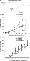
In vivo therapy of HCT15 colon tumor xenografts. Mice were divided into four groups to assess combination therapy in vivo when the diameters of tumors reached approximately 100 mm3 (▪, PBS; ▴, doxorubicin; ⧫, I-131; •, doxorubicin plus I-131). A virus mixture of Ad-shMDR1 (2 × 10⁹ PFU) and Ad-hNIS (1 × 10⁹ PFU) was administered to mice by multiple injections of virus. Ad-LacZ as a control was intratumorally injected to mice by multiple injections of virus. At 2 days after virus injection, a single dose of I-131 (55.5 MBq) was injected and doxorubicin treatment (0.5 mg kg–1) was followed at 3-day intervals for 21 days (a). Tumor-bearing mice were injected with either virus mixture (Ad-shMDR1 plus Ad-hNIS) (b) or Ad-LacZ (c) followed by treatment with PBS, doxorubicin, I-131 or doxorubicin+I-131. Tumor volume was measured using a caliper over time. Experiments were performed in triplicate, n=8 mice/group. *statistically significant.

Similar articles

Cited by

References

    1. Chung JK. Sodium iodide symporter: its role in nuclear medicine. J Nucl Med. 2002;43:1188–1200. - PubMed
    1. Cho JY. A transporter gene (sodium iodide symporter) for dual purposes in gene therapy: imaging and therapy. Curr Gene Ther. 2002;2:393–402. - PubMed
    1. Chen L, Altmann A, Mier W, Eskerski H, Leotta K, Guo L, et al. Radioiodine therapy of hepatoma using targeted transfer of the human sodium/iodide symporter gene. J Nucl Med. 2006;47:854–862. - PubMed
    1. Mandell RB, Mandell LZ, Link CJ., Jr Radioisotope concentrator gene therapy using the sodium/iodide symporter gene. Cancer Res. 1999;59:661–668. - PubMed
    1. Jeon YH, Choi Y, Kim HJ, Kim CW, Jeong JM, Lee DS, et al. Human sodium iodide symporter gene adjunctive radiotherapy to enhance the preventive effect of hMUC1 DNA vaccine. Int J Cancer. 2007;121:1593–1599. - PubMed

Publication types

MeSH terms