. 2010 Feb 23;5(2):e9380.
doi: 10.1371/journal.pone.0009380.

Myelin activates FAK/Akt/NF-kappaB pathways and provokes CR3-dependent inflammatory response in murine system

Affiliations

Myelin activates FAK/Akt/NF-kappaB pathways and provokes CR3-dependent inflammatory response in murine system

Xin Sun et al. PLoS One. .

Abstract

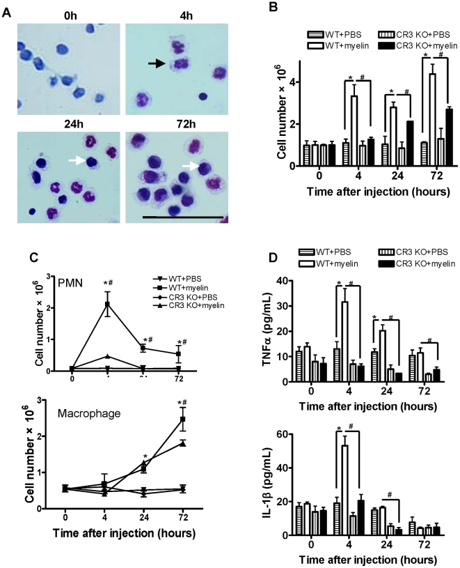
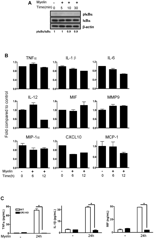
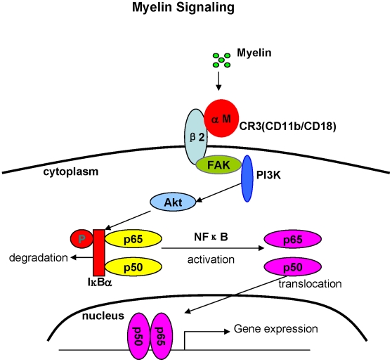
Inflammatory response following central nervous system (CNS) injury contributes to progressive neuropathology and reduction in functional recovery. Axons are sensitive to mechanical injury and toxic inflammatory mediators, which may lead to demyelination. Although it is well documented that degenerated myelin triggers undesirable inflammatory responses in autoimmune diseases such as multiple sclerosis (MS) and its animal model, experimental autoimmune encephalomyelitis (EAE), there has been very little study of the direct inflammatory consequences of damaged myelin in spinal cord injury (SCI), i.e., there is no direct evidence to show that myelin debris from injured spinal cord can trigger undesirable inflammation in vitro and in vivo. Our data showed that myelin can initiate inflammatory responses in vivo, which is complement receptor 3 (CR3)-dependent via stimulating macrophages to express pro-inflammatory molecules and down-regulates expression of anti-inflammatory cytokines. Mechanism study revealed that myelin-increased cytokine expression is through activation of FAK/PI3K/Akt/NF-kappaB signaling pathways and CR3 contributes to myelin-induced PI3K/Akt/NF-kappaB activation and cytokine production. The myelin induced inflammatory response is myelin specific as sphingomyelin (the major lipid of myelin) and myelin basic protein (MBP, one of the major proteins of myelin) are not able to activate NF-kappaB signaling pathway. In conclusion, our results demonstrate a crucial role of myelin as an endogenous inflammatory stimulus that induces pro-inflammatory responses and suggest that blocking myelin-CR3 interaction and enhancing myelin debris clearance may be effective interventions for treating SCI.


Conflict of interest statement

Competing Interests: The authors have declared that no competing interests exist.

Figures

Figure 1
Figure 1. Inflammatory effect of myelin in peritoneal cavity from WT and CR3 KO mice.
(A) Morphology of infiltrated cells in peritoneal cavity from WT mice. Neutrophils were indicated with black arrow and macrophages were indicated with white arrow. (Scale bar = 50 µm). (B) The total number of cells in peritoneal cavity. (C) Number of infiltrated neutrophils and macrophages in peritoneal cavity. (D) Levels of TNF-α and IL-1β in peritoneal lavage fluid detected by ELISA. Data were expressed as mean ± SEM. (n = 4 at each time point, *P<0.05 vs. WT + PBS; #P<0.05 vs. CR3 KO + Myelin; t-test).
Figure 2
Figure 2. Inflammatory effect of myelin in spinal cord from WT and CR3 KO mice.
(A) Neutrophils were positive for Ly-6G (green) but negative for IBA-1 (red) detected by double immunostaining. (B) Neutrophil and macrophage infiltration in spinal cord at 1 d after PBS injection detected by double immunostaining. (C) Neutrophil infiltration (Ly-6G+, green) in spinal cord at 1 d after myelin injection, along with injection region (solid line), marginal region (dashed line) and enlarged injection region. (D) The number of neutrophils at injection region and marginal region in spinal cord at 1 d after myelin or PBS injection. (E) Macrophage infiltration (IBA-1+, red) in spinal cord at 4 d after myelin injection, along with injection region (solid line), marginal region (dashed line) and enlarged injection region. (F) The number of macrophages at injection and marginal region in spinal cord at 4 d after myelin or PBS injection. Scale bar = 200 µm. Data were expressed as mean ± SEM. (n = 4 at each time point, *P<0.05 vs. WT + PBS; #P<0.05 vs. CR3 KO + myelin; t-test).
Figure 3
Figure 3. Expression of inflammatory mediators at transcriptional and translational levels in bone marrow-derived macrophages regulated by myelin.
(A) Cytokines and chemokines in the cells detected by qRT-PCR. (B) Cytokines in the supernatants of cell culture detected by ELISA. Data were expressed as mean ± SEM and similar results were obtained from three independent experiments. (n = 3. *P<0.05 vs. control; one way ANOVA with Dunnett's post-test).
Figure 4
Figure 4. Effect of myelin on NF-κB activation.
(A) Bone marrow-derived macrophages were incubated with myelin for indicated time and phosphorylation/degradation of IκB-α were assessed by western blot. (B) Effect of NF-κB inhibitor BAY 11-7082 on myelin-induced IκB-α phosphorylation and degradation. (C) Myelin-induced p65 translocation in macrophages detected by immunofluorescent staining. p65 was stained red and nuclei were stained with DAPI (blue) (Scale bar = 10 µm). (D) Sphingomyelin (25 µg/mL) had no effect on IκB-α phosphorylation and degradation macrophages. (E) MBP (1 µg/mL) had no effect on IκB-α phosphorylation and degradation in bone marrow-derived macrophages. The inhibitor was used for 30-minute pre-incubation before myelin incubation. Phosphorylation levels were normalized to non-phosphorylated total proteins. Similar results were obtained from three independent experiments.
Figure 5
Figure 5. Effect of inactivation of NF-κB on myelin-induced cytokine expression in macrophages.
(A) Macrophages were pre-treated with BAY 11-7082 for 30 minutes and then treated with myelin for 12 hours. Cytokines at transcriptional level were detected by qRT-PCR. (B) Macrophages were pre-treated with BAY 11-7082 for 30 minutes and then treated with myelin for 24 hours. Cytokines at protein level were detected by ELISA. Data were expressed as mean ± SEM and similar results were obtained from three independent experiments. (n = 3, *P<0.05 vs. control; #P<0.05 vs. BAY treatment; t-test).
Figure 6
Figure 6. Effect of myelin on NF-κB activation and cytokine expression in bone marrow-derived macrophages from CR3 KO mice.
(A) Macrophages from CR3 KO mice were treated with myelin for the indicated time and phosphorylation/degradation of IκB-α were assessed by western blot. (B) Macrophages from CR3 KO mice were treated with myelin for 6 and 12 hours. Cytokine and chemokine expression in cells at mRNA levels were detected by qRT-PCR. (C) Macrophages from WT and CR3 KO mice were treated with myelin for 24 hours and cytokines in the supernatants of cell culture were detected by ELISA. Phosphorylation levels were normalized to non-phosphorylated total proteins. Data were expressed as mean ± SEM and similar results were obtained from three independent experiments. (n = 3, *P<0.05 vs. CR3 KO + myelin; t-test).
Figure 7
Figure 7. Effect of myelin on FAK phosphorylation.
Bone marrow-derived macrophages from WT and CR3 KO mice were treated with myelin for the indicated time and phosphorylation of FAK (pFAK) was assessed by western blot. Phosphorylation levels were normalized to non-phosphorylated total proteins. Similar results were obtained from three independent experiments.
Figure 8
Figure 8. Effect of myelin on activation of PI3K/Akt pathway.
(A) Phosphorylation of p85 and Akt in bone marrow-derived macrophages from WT and CR3 KO mice treated with myelin for the indicated time. (B) Effect of PI3K inhibitor LY-294002 on myelin-induced IκB-α phosphorylation and degradation in macrophages. (C) Effect of Akt inhibitor IV on myelin-induced IκB-α phosphorylation and degradation in macrophages. (D) Effect of inhibitors of PI3K, Akt, NF-κB and PKC on myelin-induced Akt phosphorylation in macrophages. (E) Macrophages were pretreated with inhibitors of PI3K, Akt, NF-κB and PKC for 30 minutes, respectively and then incubated with myelin for 90 minutes. Myelin-induced p65 translocation in macrophages was detected by immunofluorescent staining. p65 was stained red and nuclei were stained with DAPI (blue) (Scale bar = 10 µm). All inhibitors were used for 30-minute pre-incubation before myelin incubation. Phosphorylation levels were normalized to non-phosphorylated total proteins. Similar results were obtained from three independent experiments.
Figure 9
Figure 9. Effect of inactivation of PI3K and Akt on myelin-induced cytokine expression in macrophages.
Macrophages were pre-treated with LY-294002 (A) or Akt inhibitor IV (B) for 30 minutes and then treated with myelin for 12 hours. Cytokines at transcriptional level were detected by qRT-PCR. Data were expressed as mean ± SEM and similar results were obtained from three independent experiments. (n = 3, *P<0.05 vs. control; #P<0.05 vs. inhibitor treatment; t-test).
Figure 10
Figure 10. Schematic diagram depicting the possible mechanism that myelin induces inflammatory responses.
Myelin debris generated in injured CNS or demyelinating diseases may bind to CR3 and then activate NF-κB through FAK/PI3K/Akt pathway, therefore initiating gene expression of pro-inflammatory mediators.

References

    1. Donnelly DJ, Popovich PG. Inflammation and its role in neuroprotection, axonal regeneration and functional recovery after spinal cord injury. Exp Neurol. 2008;209:378–388. - PMC - PubMed
    1. Hausmann ON. Post-traumatic inflammation following spinal cord injury. Spinal Cord. 2003;41:369–378. - PubMed
    1. Popovich PG, Wei P, Stokes BT. Cellular inflammatory response after spinal cord injury in Sprague-Dawley and Lewis rats. J Comp Neurol. 1997;377:443–464. - PubMed
    1. Pineau I, Lacroix S. Proinflammatory cytokine synthesis in the injured mouse spinal cord: multiphasic expression pattern and identification of the cell types involved. J Comp Neurol. 2007;500:267–285. - PubMed
    1. Profyris C, Cheema SS, Zang D, Azari MF, Boyle K, et al. Degenerative and regenerative mechanisms governing spinal cord injury. Neurobiol Dis. 2004;15:415–436. - PubMed

Publication types

MeSH terms