. 2010 May;53(5):989-1000.
doi: 10.1007/s00125-010-1677-0. Epub 2010 Feb 26.

Hyperglycaemia-induced impairment of endothelium-dependent vasorelaxation in rat mesenteric arteries is mediated by intracellular methylglyoxal levels in a pathway dependent on oxidative stress

Affiliations

Hyperglycaemia-induced impairment of endothelium-dependent vasorelaxation in rat mesenteric arteries is mediated by intracellular methylglyoxal levels in a pathway dependent on oxidative stress

O Brouwers et al. Diabetologia. 2010 May.

Abstract

Aims/hypothesis: Impaired nitric oxide (NO)-dependent vasorelaxation plays a key role in the development of diabetic vascular complications. We investigated the effect of hyperglycaemia on impaired vasoreactivity and a putative role therein of the AGE precursor methylglyoxal.

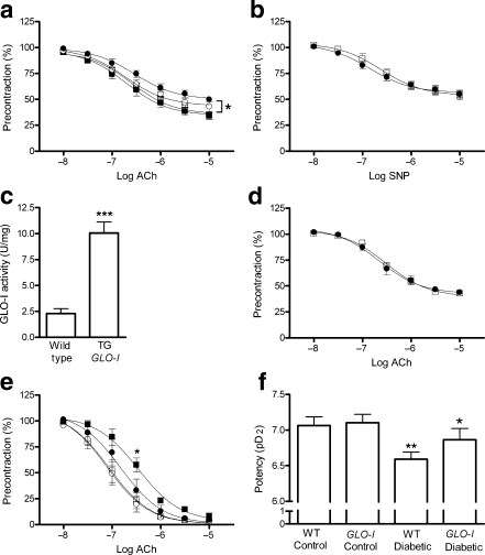
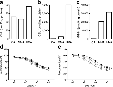
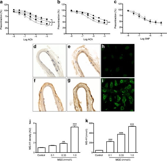
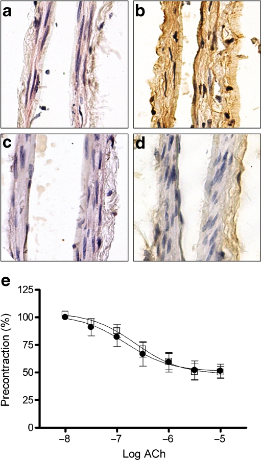
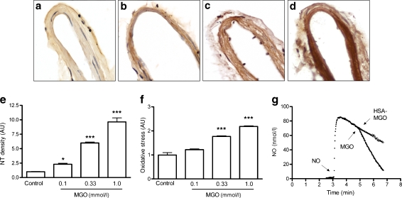
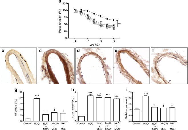
Methods: The effects of high glucose and methylglyoxal on NO-dependent vasorelaxation in isolated rat mesenteric arteries from wild-type and transgenic glyoxalase (GLO)-I (also known as GLO1) rats, i.e. the enzyme detoxifying methylglyoxal, were recorded in a wire myograph. AGE formation of the major methylglyoxal-adduct 5-hydro-5-methylimidazolone (MG-H1) was detected with an antibody against MG-H1 and quantified with ultra-performance liquid chromatography (tandem) mass spectrometry. Reactive oxygen species formation was measured with a 5-(and-6)-chloromethyl-2'7'-dichlorodihydrofluorescein diacetate acetyl ester probe and by immunohistochemistry with an antibody against nitrotyrosine.

Results: High glucose and methylglyoxal exposure of mesenteric arteries significantly reduced the efficacy of NO-dependent vasorelaxation (p < 0.05). This impairment was not observed in mesenteric arteries of GLO-I transgenic rats indicating a specific intracellular methylglyoxal effect. The diabetes-induced impaired potency (pD(2)) in mesenteric arteries of wild-type rats was significantly improved by GLO-I overexpression (p < 0.05). Methylglyoxal-modified albumin did not affect NO-dependent vasorelaxation, while under the same conditions the receptor for AGE ligand S100b did (p < 0.05). Methylglyoxal treatment of arteries increased intracellular staining of MG-H1 in endothelial cells and adventitia by fivefold accompanied by an eightfold increase in the oxidative stress marker nitrotyrosine. Antioxidant pre-incubation prevented methylglyoxal-induced impairment of vasoreactivity.

Conclusions/interpretation: These data show that hyperglycaemia-induced impairment of endothelium-dependent vasorelaxation is mediated by increased intracellular methylglyoxal levels in a pathway dependent on oxidative stress.


Figures

Fig. 1
Fig. 1
Effect of high glucose concentration on acetylcholine (ACh)-induced NO-mediated endothelium-dependent relaxation in isolated rat mesenteric arteries. a Rat mesenteric arteries were mounted in a myograph and incubated with or without high glucose concentrations for 2 h. Acetylcholine-induced NO-mediated endothelium-dependent relaxation was impaired in a concentration-dependent manner during incubation with 30 mmol/l glucose (white circles) or 40 mmol/l glucose (black circles) compared with control (5 mmol/l glucose, white squares). The 40 mmol/l glucose incubation resulted in a statistically significant shift of the dose–response curve (*p < 0.05 vs control group). Incubation with 35 mmol/l mannitol (black squares) as an osmotic control did not impair NO-mediated endothelium-dependent relaxation (n = 6). b SNP-induced NO-mediated endothelium-independent vasorelaxation during incubation without (white squares) or with 40 mmol/l glucose (black circles) for 2 h did not differ (n = 6). c GLO-I activity in mesenteric arteries homogenates of GLO-I transgenic (TG) rats was significantly elevated compared with mesenteric arteries homogenates of wild-type rats (***p < 0.001, n = 4). d In mesenteric arteries of GLO-I transgenic rats, acetylcholine-induced NO-mediated endothelium-dependent relaxation was not impaired during incubation with 40 mmol/l glucose (black circles) for 2 h compared with control incubations (5 mmol/l glucose, white squares, n = 5). e Acetylcholine-induced vasorelaxation was significantly impaired in wild-type diabetic rats (black squares; *p < 0.05) compared with the controls (wild-type control, white squares; transgenic control, white circles). GLO-I overexpression improved this relaxation (black circles; n = 8). f The decreased potency in wild-type (WT) diabetic rats (**p < 0.01) was significantly improved by GLO-I overexpression (*p < 0.05)
Fig. 2
Fig. 2
Effect of methylglyoxal-modified albumin and S100b on acetylcholine-induced NO-mediated endothelium-dependent relaxation in isolated rat mesenteric arteries. a–c Characteristics of methylglyoxal-modified albumin (MMA). Control albumin (CA) was exposed to methylglyoxal (0.5 and 10 mmol/l) for 2 days to prepare minimally modified methylglyoxal-albumin and highly modified methylglyoxal-albumin (HMA). The amount of CML (a), CEL (b) and MG-H1 (c) was determined by UPLC-MSMS analysis. d Acetylcholine (ACh)-induced NO-mediated endothelium-dependent relaxation during incubation with albumin (black squares), minimally modified methylglyoxal-albumin (white circles) and highly modified methylglyoxal-albumin (black circles) did not statistically differ from the control group (white squares); n = 4. e Incubation of mesenteric arteries with 20 (white circles) or 40 µg/ml (black circles) of the RAGE ligand S100b resulted in concentration-dependent impairment of acetylcholine-induced NO-mediated endothelium-dependent relaxation. The 40 µg/ml incubation resulted in a statistically significant shift of the dose–response curve; *p < 0.05 vs control group, n = 4
Fig. 3
Fig. 3
Effect of methylglyoxal on vascular reactivity and intracellular MG-H1 formation. a Incubation of isolated mesenteric arteries for 1 h with 0.1 mmol/l (black squares), 0.33 mmol/l (white circles) and 1.0 mmol/l (black circles) methylglyoxal resulted in impairment of acetylcholine (ACh)-induced NO-mediated endothelium-dependent relaxation compared with the control group (white squares); n = 5 for all conditions. Incubation with 0.33 mmol/l and 1.0 mmol/l methylglyoxal resulted in a statistically significant shift of the dose–response curve (*p < 0.05 and ***p < 0.001 respectively vs control group; n = 5). b Incubation of isolated mesenteric arteries for 2 h with 10 µmol/l (black squares) and 50 µmol/l (black circles) methylglyoxal resulted in impairment of acetylcholine-induced NO-mediated endothelium-dependent relaxation compared with the control group (white squares); n = 4 for all conditions. Incubation with 50 µmol/l methylglyoxal resulted in a statistically significant shift of the dose–response curve (**p < 0.01 vs control group; n = 4). c SNP-induced NO-mediated endothelium-independent vasorelaxation during incubation without (white squares) or with 1.0 mmol/l methylglyoxal (black circles) for 1 h did not differ (n = 4). d–i Localisation of MG-H1 in adventitia and endothelial cells of mesenteric arteries not incubated (d) or incubated with 0.1 mmol/l (e), 0.33 mmol/l (f) or 1.0 mmol/l (g) methylglyoxal for 1 h. Intracellular localisation of MG-H1 was also determined by confocal laser scanning microscopy in cultured endothelial cells (ECRF24) incubated without (h) or with 1.0 mmol/l methylglyoxal (i) for 1 h. j Quantification of MG-H1 staining in arteries exposed to methylglyoxal (n = 4). k Concentration-dependent increase of MG-H1 levels in protein lysates of mesenteric arteries not incubated (Control) or incubated with 0.1, 0.33 or 1.0 mmol/l methylglyoxal (MGO) for 1 h as determined by UPLC-MSMS (n = 3). j, k **p < 0.01 and ***p < 0.001 vs control group. AU, arbitrary units
Fig. 4
Fig. 4
Effect of GLO-I overexpression on methylglyoxal (MGO)-induced formation of the major methylglyoxal-adduct MG-H1 and on vascular reactivity. a–d Compared with MG-H1 staining in mesenteric arteries of wild-type rats incubated without (a) and with 1.0 mmol/l methylglyoxal (b), incubation for 1 h with 1.0 mmol/l methylglyoxal did not lead to intracellular MG-H1 formation in GLO-I transgenic rats incubated without (c) and with 1.0 mmol/l methylglyoxal (d). e In mesenteric arteries of GLO-I transgenic rats, acetylcholine (ACh)-induced NO-mediated endothelium-dependent relaxation was not impaired during incubation with 1.0 mmol/l methylglyoxal (black circles) for 1 h compared with control incubation (white squares) (n = 4)
Fig. 5
Fig. 5
Methylglyoxal (MGO)-induced oxidative stress formation in isolated mesenteric arteries and cultured endothelial cells, and direct quenching of NO. a–d Representive staining of nitrotyrosine (NT) in mesenteric resistance arteries not incubated (a) or incubated for 1 h with 0.1 mmol/l (b), 0.33 mmol/l (c) or 1.0 mmol/l (d) methylglyoxal. e Quantification of NT staining in methylglyoxal-incubated arteries (n = 4). f Oxidative stress formation in endothelial cells as measured with a CM-H2DCFDA probe during treatment for 1 h with methylglyoxal as shown; n = 3. g Direct effect of methylglyoxal (MGO) and methylglyoxal-modified human albumin (HAS-MGO) on NO as measured with an Iso-NO meter in deoxygenated buffer. *p < 0.05 and ***p < 0.001 vs control group. AU, arbitrary units
Fig. 6
Fig. 6
Effect of antioxidants on the effect of methylglyoxal on vascular reactivity and oxidative stress formation. a Acetylcholine (ACh)-induced NO-mediated endothelium-dependent relaxation during incubation without (white squares) or with 1.0 mmol/l methylglyoxal (black circles) for 1 h with or without pre-incubation for 20 min with the antioxidants NAC (1.0 mmol/l) (white diamonds), EUK-134 (1 µmol/l) (black squares) or Mn(III)TMP (1 µmol/l) (white circles). Treatment of mesenteric arteries with 1.0 mmol/l methylglyoxal in the absence of antioxidant resulted in a significant shift in the dose-response curve compared with the control group (***p < 0.001). All antioxidant pretreatments prevented methylglyoxal-induced impairment of reactivity (n = 5). b–f Nitrotyrosine staining of arteries incubated without (b), or with 1.0 mmol/l methylglyoxal (c) and after pre-incubation with 1.0 mmol/l NAC (d), 1 µmol/l EUK-134 (e) or 1 µmol/l Mn(III)TMP (f). g Quantification of NT staining in mesenteric arteries after treatment with 1.0 mmol/l methylglyoxal (MGO) without or with pre-incubation NAC, EUK-134 or Mn(III)TMP (n = 4). h Quantification of MG-H1 staining in mesenteric arteries after treatment with 1.0 mmol/l methylglyoxal without or with pre-incubation at the same concentrations of NAC, EUK-134 or Mn(III)TMP. i Oxidative stress formation measured with a CM-H2DCFDA probe in ECRF24 cells after treatment with 1.0 mmol/l methylglyoxal without or with pre-incubation at 100 µmol/l NAC, 10 nmol/l EUK-134 and 10 nmol/l Mn(III)TMP (n = 3). g–i ***p < 0.001 vs control group; †p < 0.001 vs methylglyoxal group. AU, arbitrary units

Similar articles

Cited by

References

    1. Schalkwijk CG, Stehouwer CD. Vascular complications in diabetes mellitus: the role of endothelial dysfunction. Clin Sci (Lond) 2005;109:143–159. doi: 10.1042/CS20050025. - DOI - PubMed
    1. Steinberg HO, Chaker H, Leaming R, Johnson A, Brechtel G, Baron AD. Obesity/insulin resistance is associated with endothelial dysfunction. Implications for the syndrome of insulin resistance. J Clin Invest. 1996;97:2601–2610. doi: 10.1172/JCI118709. - DOI - PMC - PubMed
    1. Makimattila S, Virkamaki A, Groop PH, et al. Chronic hyperglycemia impairs endothelial function and insulin sensitivity via different mechanisms in insulin-dependent diabetes mellitus. Circulation. 1996;94:1276–1282. - PubMed
    1. Diederich D, Skopec J, Diederich A, Dai FX. Endothelial dysfunction in mesenteric resistance arteries of diabetic rats: role of free radicals. Am J Physiol. 1994;266:H1153–H1161. - PubMed
    1. Pflueger AC, Osswald H, Knox FG. Adenosine-induced renal vasoconstriction in diabetes mellitus rats: role of nitric oxide. Am J Physiol. 1999;276:F340–F346. - PubMed

Publication types

MeSH terms