

The fate of porous hydroxyapatite granules used in facial skeletal augmentation
- PMID: 20186415
- PMCID: PMC2906722
- DOI: 10.1007/s00266-010-9473-2
The fate of porous hydroxyapatite granules used in facial skeletal augmentation
Abstract
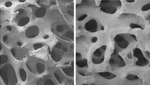
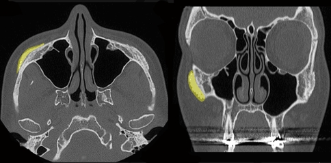
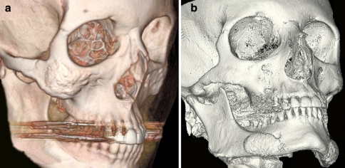
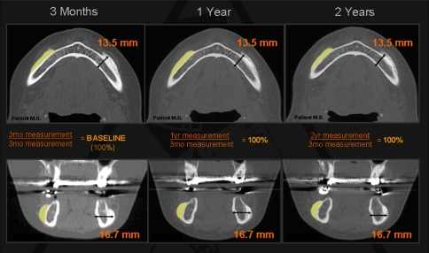
Facial appearance is largely determined by the morphology of the underlying skeleton. Hydroxyapatite is one of several materials available to enhance projection of the facial skeleton. This study evaluated the long-term maintenance of augmented bony projection when porous hydroxyapatite granules are used on the facial skeleton. Ten female patients aged 28-58 years were studied following aesthetic augmentation of the facial skeleton at 24 sites using porous hydroxyapatite granules. Postoperative CT scans at 3 months served as the baseline measurement and compared with scans taken at 1 and 2 years, with the thickness of the hydroxyapatite measured in axial and coronal planes. Thickness of original bone plus overlay of hydroxyapatite, thickness of the overlying soft tissue, and the overall projection (bone plus soft tissue) were recorded. It was found that 99.7% of the hydroxyapatite was maintained at 2 years, with no statistical difference (t test) from the baseline measurement. The overall projection (bony and soft tissue) was maintained as there was no evidence of native bone resorption or soft tissue atrophy. Radiographic results confirmed that the use of porous hydroxyapatite granules for enhancement of the facial skeleton is not only a predictable procedure, but maintains full bony projection at 2 years.
Figures
Similar articles
-
Augmentation of the craniomaxillofacial region using porous hydroxyapatite granules.Plast Reconstr Surg. 2003 May;111(6):1808-17. doi: 10.1097/01.PRS.0000055432.20074.93. Plast Reconstr Surg. 2003. PMID: 12711940
-
Augmentation of the craniofacial skeleton with porous hydroxyapatite granules.Plast Reconstr Surg. 1993 Jan;91(1):15-22; discussion 23-6. Plast Reconstr Surg. 1993. PMID: 8380106
-
Biologic Behavior of Hydroxyapatite Used in Facial Augmentation.Aesthetic Plast Surg. 2017 Feb;41(1):179-184. doi: 10.1007/s00266-016-0707-9. Epub 2016 Dec 23. Aesthetic Plast Surg. 2017. PMID: 28008459
-
Skeletal volume enhancement: implants and osteotomies.Curr Opin Otolaryngol Head Neck Surg. 2004 Aug;12(4):349-56. doi: 10.1097/01.moo.0000130576.04818.55. Curr Opin Otolaryngol Head Neck Surg. 2004. PMID: 15252260 Review.
-
The versatility of hydroxyapatite blocks in maxillofacial surgery.Br J Oral Maxillofac Surg. 1987 Dec;25(6):452-64. doi: 10.1016/0266-4356(87)90137-9. Br J Oral Maxillofac Surg. 1987. PMID: 2825758 Review.
Cited by
-
Changes in the facial skeleton with aging: implications and clinical applications in facial rejuvenation.Aesthetic Plast Surg. 2012 Aug;36(4):753-60. doi: 10.1007/s00266-012-9904-3. Epub 2012 May 12. Aesthetic Plast Surg. 2012. PMID: 22580543 Free PMC article. Review.
-
Early Odontogenic Differentiation of Dental Pulp Stem Cells Treated with Nanohydroxyapatite-Silica-Glass Ionomer Cement.Polymers (Basel). 2020 Sep 17;12(9):2125. doi: 10.3390/polym12092125. Polymers (Basel). 2020. PMID: 32957636 Free PMC article.
-
Safety and Efficacy of Midface Augmentation Using Bio-Oss Bone Powder and Bio-Gide Collagen Membrane in Asians.J Clin Med. 2023 Jan 26;12(3):959. doi: 10.3390/jcm12030959. J Clin Med. 2023. PMID: 36769607 Free PMC article.
-
Biomimetic Approaches in Clinical Endodontics.Biomimetics (Basel). 2022 Dec 6;7(4):229. doi: 10.3390/biomimetics7040229. Biomimetics (Basel). 2022. PMID: 36546929 Free PMC article. Review.
-
Dual Modification of Porous Ca-P/PLA Composites with APTES and Alendronate Improves Their Mechanical Strength and Cytobiocompatibility towards Human Osteoblasts.Int J Mol Sci. 2022 Nov 18;23(22):14315. doi: 10.3390/ijms232214315. Int J Mol Sci. 2022. PMID: 36430791 Free PMC article.
References
-
- Rees TD, Wood Smith D. Cosmetic facial surgery. 1. Philadelphia: Saunders; 1973.
-
- Rosen HM. Treatment planning: aesthetic goals. New York: Springer; 1999.
MeSH terms
Substances
LinkOut - more resources
Full Text Sources
Medical
