. 2010 Jun;67(11):1919-27.
doi: 10.1007/s00018-010-0302-1. Epub 2010 Feb 26.

A 629RKLKK633 motif in the hinge region controls the androgen receptor at multiple levels

Affiliations

A 629RKLKK633 motif in the hinge region controls the androgen receptor at multiple levels

Tamzin M Tanner et al. Cell Mol Life Sci. 2010 Jun.

Abstract

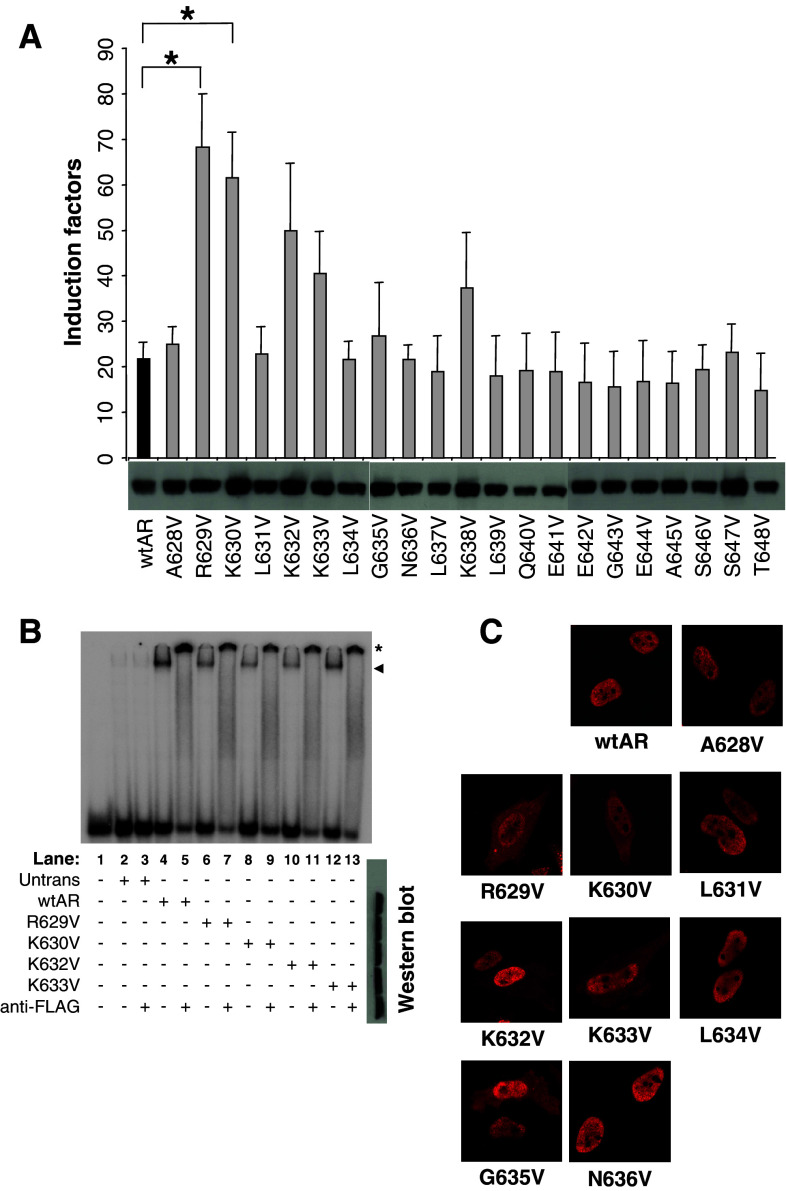
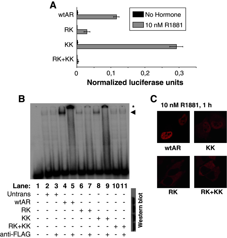
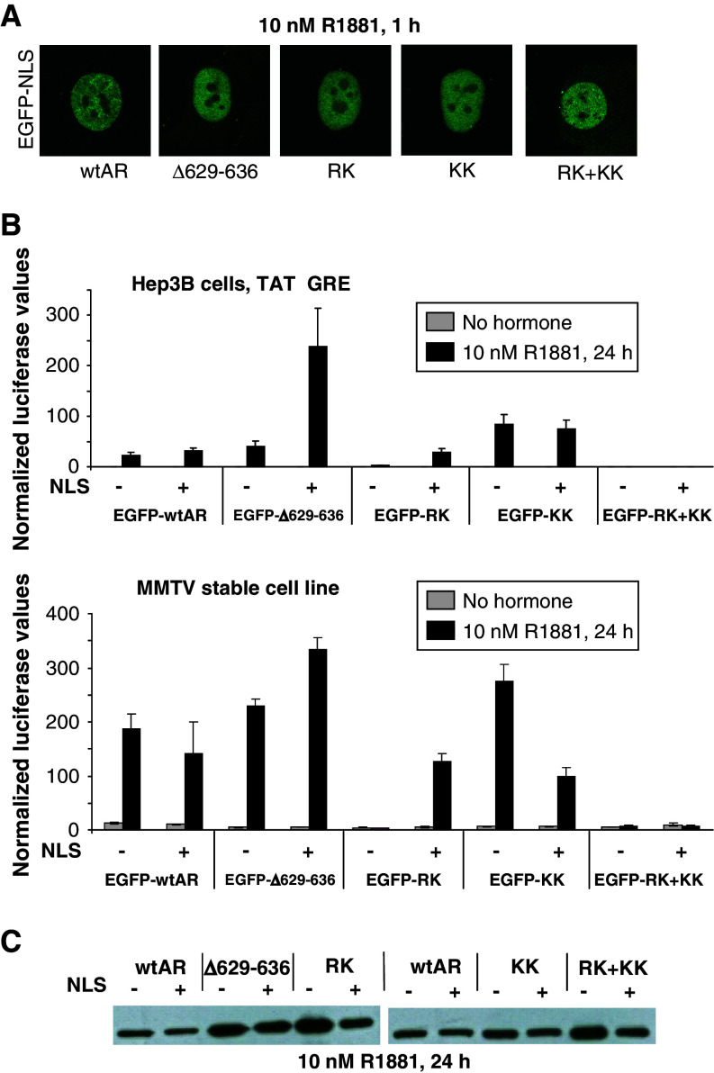
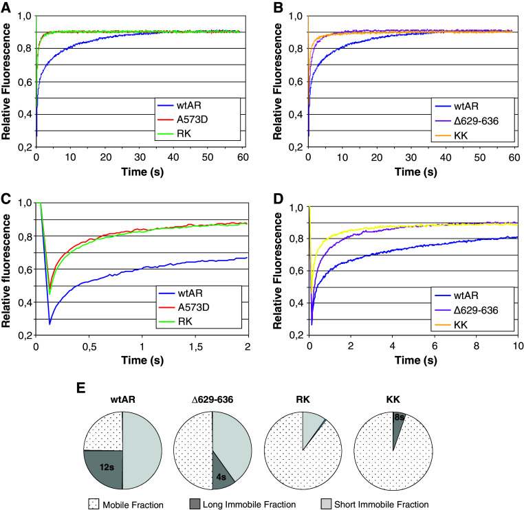
The androgen receptor protein has specific domains involved in DNA binding, ligand binding, and transactivation, whose activities need to be integrated during transcription activation. The hinge region, more particular a (629)RKLKK(633) motif, seems to play a crucial role in this process. Indeed, although the motif is not part of the DNA-binding domain, its positive residues are involved in optimal DNA binding and nuclear translocation as shown by mutation analysis. When the mutated ARs are forced into the nucleus, however, the residues seem to play different roles in transactivation. Moreover, we show by FRAP analysis that during activation, the AR is distributed in the nucleus in a mobile and two immobile fractions, and that mutations in the (629)RKLKK(633) motif affect the distribution of the AR over these three intranuclear fractions. Taken together, the (629)RKLKK(633) motif is a multifunctional motif that integrates nuclear localization, receptor stability, DNA binding, transactivation potential and intranuclear mobility.


Figures

Fig. 1
Fig. 1
Substitution analysis of the AR hinge region. a Functional analysis. A TAT-GRE-based reporter was co-transfected in HeLa cells with AR expression plasmids as indicated. Results are shown as 10 nM R1881 induction factors ± SEM. *p < 0.05 versus wtAR as determined by the Student’s t-test. Western-blot data for each mutant is given below the X-axis. b Electromobility shift assays. The labeled TAT-GRE probe was incubated with cell extracts containing the indicated mutant AR. The shifted complexes are indicated with an arrowhead. The supershifts, which are indicated with an asterisk, were obtained by adding an anti-flag antibody. Western blots of the extracts are shown at the right. c Nuclear translocation studies. HeLa cells were transfected with the indicated AR expression vectors, stimulated, fixed, and immunostained. Cellular localization of the expressed AR proteins was analyzed by fluorescence microscopy
Fig. 2
Fig. 2
The effect of duplicate or quadruplicate mutations in the 629RKLKK633 motif. a Functional analysis. A TAT-GRE-based reporter was co-transfected in HeLa cells with AR expression plasmids as indicated. Results are shown as normalized luciferase units. Western-blot data for each mutant is given below the X-axis. b Electrophoretic mobility shift assays. The labeled TAT-GRE probe was incubated with cell extracts in which the mutated ARs have been over-expressed. The shifted complexes are indicated with an arrowhead. The supershifts, which are indicated with an asterisk, were obtained by adding an anti-flag antibody. Western blots of the extracts are given at the right. c Nuclear translocation studies. HeLa cells were transfected with the indicated AR expression vectors, stimulated, fixed, and immunostained. Cellular localization of the expressed AR proteins was analyzed by fluorescence microscopy
Fig. 3
Fig. 3
Activity of hinge region mutants when localized in the nucleus. a Nuclear localization study. Confocal images of Hep3B cells stably expressing EGFP-NLS-AR proteins. b Functional analysis. Hep3B cells or the MMTV stable cell line were transiently transfected with the indicated expression vector. For the Hep3B cells, the TAT-GRE-luc reporter was cotransfected. Results are shown as normalized luciferase values ± SEM. c Western blot. The indicated receptor proteins fused to EGFP plus or minus the SV40-NLS were expressed in HeLa cells and detected with an anti-flag antibody
Fig. 4
Fig. 4
Intranuclear distribution and mobility of EGFP-NLS-AR proteins as measured by FRAP. ad Fitted curves of experimental FRAP data. FRAP data of wtAR is plotted on all curves for comparative purposes. Three independent experiments were performed and data represent the mean of at least 45 cells. e Schematic representation of analysis of FRAP data. FRAP data fitted best with simulated curves with indicated parameters for immobile fractions and residence times. The percentage of molecules residing within the long immobile fraction (LIF) and short immobile fraction (SIF) are quantified in the pie charts. The remaining molecules were assumed to all reside in the freely diffusing mobile fraction. Residence time characterizes the mean immobilization of individual ARs in seconds. The calculated residence time for the various LIFs is indicated on the pie charts

Similar articles

Cited by

References

    1. Métivier R, Reid G, Gannon F. Transcription in four dimensions: nuclear receptor-directed initiation of gene expression. EMBO reports. 2006;7:161–167. doi: 10.1038/sj.embor.7400626. - DOI - PMC - PubMed
    1. Xu P, Liu Y, Shan S, Kong Y, Zhou Q, Li M, Ding J, Xie Y, Wang Y. Molecular mechanism for the potentiation of the transcriptional activity of human liver receptor homolog 1 by steroid receptor coactivator-1. Mol Endocrinol. 2004;18:1887–1905. doi: 10.1210/me.2003-0334. - DOI - PubMed
    1. Iordanidou P, Aggelidou E, Demetriades C, Hadzopoulou-Cladaras M. Distinct amino acid residues may be involved in coactivator and ligand interactions in hepatocyte nuclear factor-4α. J Biol Chem. 2005;280:21810–21819. doi: 10.1074/jbc.M501221200. - DOI - PubMed
    1. Chandra V, Huang P, Hamuro Y, Raghuram S, Wang Y, Burris TP, Rastinejad F. Structure of the intact PPAR-γ-RXR-α nuclear receptor complex on DNA. Nature. 2008;456:350–356. doi: 10.1038/nature07413. - DOI - PMC - PubMed
    1. Hong W, Baniahmad A, Liu Y, Li H. Bag-1 M is a component of the in vivo DNA-glucocorticoid receptor complex at hormone-regulated promoter. J Mol Biol. 2008;384:22–30. doi: 10.1016/j.jmb.2008.09.010. - DOI - PubMed

Publication types

MeSH terms