. 2010 Sep;15(5):619-29.
doi: 10.1007/s12192-010-0174-1. Epub 2010 Feb 26.

Ero1alpha requires oxidizing and normoxic conditions to localize to the mitochondria-associated membrane (MAM)

Affiliations

Ero1alpha requires oxidizing and normoxic conditions to localize to the mitochondria-associated membrane (MAM)

Susanna Y Gilady et al. Cell Stress Chaperones. 2010 Sep.

Abstract

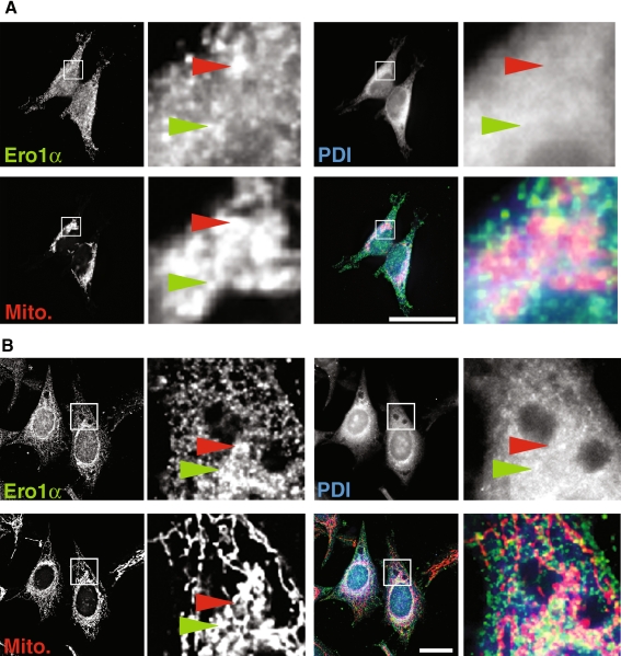
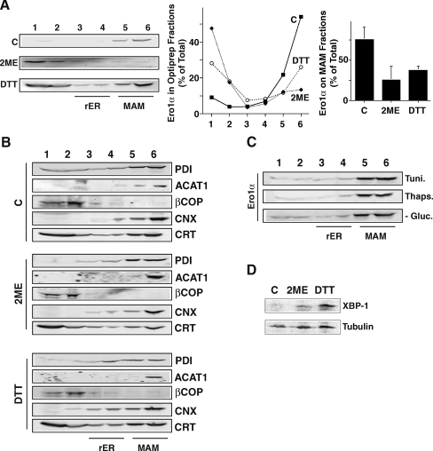
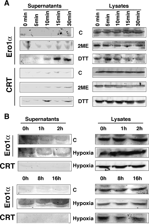
Protein secretion from the endoplasmic reticulum (ER) requires the enzymatic activity of chaperones and oxidoreductases that fold polypeptides and form disulfide bonds within newly synthesized proteins. The best-characterized ER redox relay depends on the transfer of oxidizing equivalents from molecular oxygen through ER oxidoreductin 1 (Ero1) and protein disulfide isomerase to nascent polypeptides. The formation of disulfide bonds is, however, not the sole function of ER oxidoreductases, which are also important regulators of ER calcium homeostasis. Given the role of human Ero1alpha in the regulation of the calcium release by inositol 1,4,5-trisphosphate receptors during the onset of apoptosis, we hypothesized that Ero1alpha may have a redox-sensitive localization to specific domains of the ER. Our results show that within the ER, Ero1alpha is almost exclusively found on the mitochondria-associated membrane (MAM). The localization of Ero1alpha on the MAM is dependent on oxidizing conditions within the ER. Chemical reduction of the ER environment, but not ER stress in general leads to release of Ero1alpha from the MAM. In addition, the correct localization of Ero1alpha to the MAM also requires normoxic conditions, but not ongoing oxidative phosphorylation.


Figures

Fig. 1
Fig. 1
Ero1α co-fractionates with the MAM in HEK 293 and HeLa cells. a Ero1α fractionates into heavy membranes. Membranes from HEK 293 cells were fractionated into low (HM) and high speed pellets (LM), which were analyzed by Western blotting for complex 2 (mitochondria), ACAT1 (MAM), calnexin (rER/MAM), PDI (all ER), and Ero1α. b Ero1α distribution between mitochondria and the MAM. HEK 293 homogenates were fractionated into cytosol (Cyt.), microsomes (Micro), crude mitochondria (MC), purified mitochondria (MP), and MAM according to Materials and methods. Marker proteins indicate mitochondria (complex 2), MAM (ACAT1), and ER membranes (PDI and calnexin). c Ero1α distribution within ER domains. HeLa and HEK 293 cell homogenates were fractionated on a discontinuous 10–30% Optiprep gradient. Marker proteins indicate mitochondria (complex 2), MAM (ACAT1, calnexin), pan-ER (PDI), and Ero1α. Lane numbers refer to the Optiprep fractions. The localization of Ero1α from three independent experiments is quantified in Fig. 3a. d Presence of Ero1α oxidative forms on domains of the secretory pathway. HEK293 homogenates were fractionated and analyzed under non-reducing conditions. Ero1α was detected by Western blot. e Analysis of the ER domain distribution of selected components of the ER protein folding machinery and the Golgi complex. HEK 293 cell homogenates were fractionated on a discontinuous 10–30% Optiprep gradient. eIF2α indicates the position of the rER (fractions 3 and 4), β-COP indicates the cis-Golgi (fraction 2), ACAT1 indicates the MAM (fractions 5 and 6). Calreticulin (CRT), BiP/GRP78 (BiP), ERp44, and ERp57 were analyzed for their intra-ER location
Fig. 2
Fig. 2
Intracellular localization of Ero1α to the vicinity of mitochondria. a HEK 293 cells were grown on coverslips for 24 h and processed for immunofluorescence microscopy. Ero1α was detected with a rabbit polyclonal antibody, PDI with a mouse monoclonal antiserum and mitochondria were visualized with Mitotracker. Immunofluorescence images were acquired and deconvolved. Inserts show a magnified area, indicated by white frames on the bigger pictures. The red arrowheads point out Ero1α/mitochondria overlap, whereas the green arrowheads point out triple PDI/Ero1α/mitochondria overlap. Scale bar = 25 μm. A representative image is shown. b HeLa cells were processed and imaged as in (a)
Fig. 3
Fig. 3
Ero1α MAM retention is redox-sensitive. a Optiprep fractionation of HEK 293 control cells and cells treated for 2 h with 1 mM 2ME and 1 mM DTT. Three independent experiments were probed for Ero1α and quantified by LICOR. The individual amounts of Ero1α per fraction are plotted in the middle graph (error bars omitted for clarity, but shown on the right panel). Right panel: the amount of Ero1α on the MAM was determined as the sum of signal found within fractions 5 and 6 (n = 3). b Optiprep fractionation of HEK 293 cells as in a. Western blots were probed for calnexin (CNX) and calreticulin (CRT). c Optiprep fractionation of HEK 293 cells treated with the ER stressors tunicamycin (Tuni., 10 μM), thapsigargin (Thaps., 1 μM), and in medium depleted of glucose, but supplemented with galactose (-Gluc., see Materials and methods). d Protein levels of the ER stress associated transcription factor XBP-1 after 2 h treatment with reducing agents. Loading equalized using α-tubulin as a loading control
Fig. 4
Fig. 4
Ero1α MAM retention is oxygen-sensitive. a Analysis of the Ero1α oxidation state when incubated under hypoxic conditions (1% oxygen) for the indicated times. Lines point to fully reduced Ero1α (red) and the two oxidized forms OX1 and OX2. B. Optiprep fractionation of HEK 293 control cells and cells incubated for the indicated times (in minutes and hours, see numbers on the left) in hypoxic environment (1% oxygen). Three independent experiments were probed for Ero1α and quantified by LICOR. The individual amounts of Ero1α per fraction at the 1 and 16 h time points are plotted in the middle graph (error bars omitted for clarity, but shown on the right panel). Right panel: the amount of Ero1α on the MAM was determined as the sum of signal found within fractions 5 and 6 (n = 3). c Immunofluorescence co-localization of Ero1α and βCOP at 16 h of hypoxia. HEK 293 cells were grown on coverslips for 24 h, followed by incubation in hypoxia for 16 h and processed for immunofluorescence microscopy. Ero1α was detected with a rabbit polyclonal antibody and βCOP with a mouse monoclonal antiserum. Immunofluorescence images were acquired and deconvolved. Inserts show a magnified area, indicated by white frames on the bigger pictures. Scale bar = 25 μm. A representative image is shown. Partial overlap is seen in the magnified area, but also on numerous spots elsewhere. d Optiprep fractionation of HEK 293 cells as in (a). Western blots were probed for calnexin (CNX) and BiP/GRP78 (BiP). e Optiprep fractionation of HEK 293 cells treated for 2 h with the mitochondrial stressors rotenone (1 μM), oligomycin (5 μM), and antimycin (100 μM). f HIF1α protein levels after 2 h in hypoxic environment (Hyp, 1% oxygen). Loading equalized using actin as a loading control
Fig. 5
Fig. 5
Disruption of cellular redox and oxygen supply leads to Ero1α secretion. a Ero1α content of cellular growth medium (supernatants) treated for the indicated times with the indicated reducing agents (1 mM 2ME, 1 mM DTT). Ero1α secretion, detected by the sharp increase in the growth medium (supernatants), is observed after 15 min. b Ero1α content of cellular growth medium (supernatants) incubated for the indicated times in hypoxic atmosphere. Ero1α secretion is observed after 16 h. For both a and b, calreticulin (CRT) serves as a secretion control

Similar articles

Cited by

References

    1. Anelli T, Alessio M, Mezghrani A, Simmen T, Talamo F, Bachi A, Sitia R. ERp44, a novel endoplasmic reticulum folding assistant of the thioredoxin family. EMBO J. 2002;21:835–844. doi: 10.1093/emboj/21.4.835. - DOI - PMC - PubMed
    1. Anelli T, Alessio M, Bachi A, Bergamelli L, Bertoli G, Camerini S, Mezghrani A, Ruffato E, Simmen T, Sitia R. Thiol-mediated protein retention in the endoplasmic reticulum: the role of ERp44. EMBO J. 2003;22:5015–5022. doi: 10.1093/emboj/cdg491. - DOI - PMC - PubMed
    1. Benham AM, Cabibbo A, Fassio A, Bulleid N, Sitia R, Braakman I. The CXXCXXC motif determines the folding, structure and stability of human Ero1-L alpha. Embo J. 2000;19:4493–4502. doi: 10.1093/emboj/19.17.4493. - DOI - PMC - PubMed
    1. Cabibbo A, Pagani M, Fabbri M, Rocchi M, Farmery MR, Bulleid NJ, Sitia R. ERO1-L, a human protein that favors disulfide bond formation in the endoplasmic reticulum. J Biol Chem. 2000;275:4827–4833. doi: 10.1074/jbc.275.7.4827. - DOI - PubMed
    1. Camacho P, Lechleiter JD. Calreticulin inhibits repetitive intracellular Ca2+ waves. Cell. 1995;82:765–771. doi: 10.1016/0092-8674(95)90473-5. - DOI - PubMed

Publication types