Comparative Study
. 2010 Apr;46(3-4):247-58.
doi: 10.1007/s11626-010-9297-z. Epub 2010 Feb 26.

Comparison of defined culture systems for feeder cell free propagation of human embryonic stem cells

Affiliations
Comparative Study

Comparison of defined culture systems for feeder cell free propagation of human embryonic stem cells

International Stem Cell Initiative Consortium et al. In Vitro Cell Dev Biol Anim. 2010 Apr.

Abstract

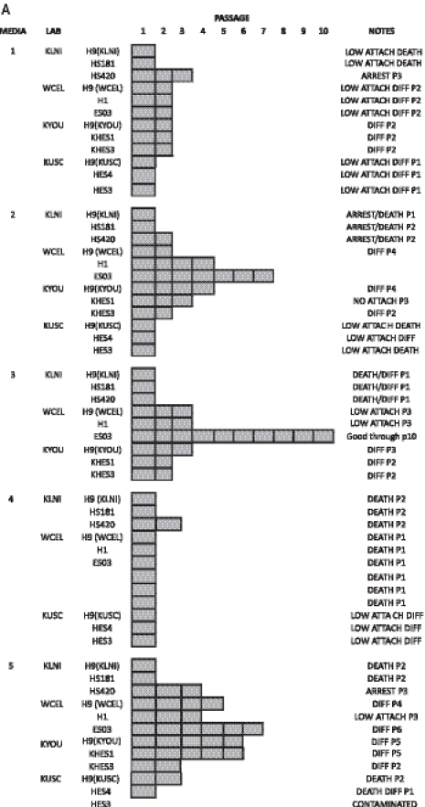
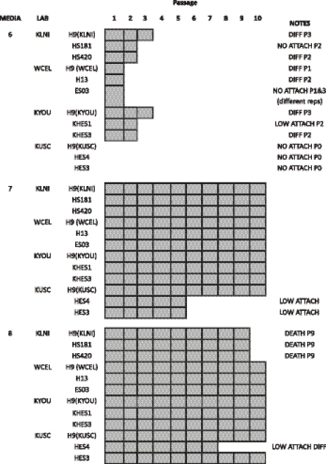
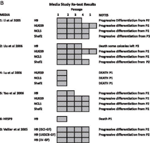
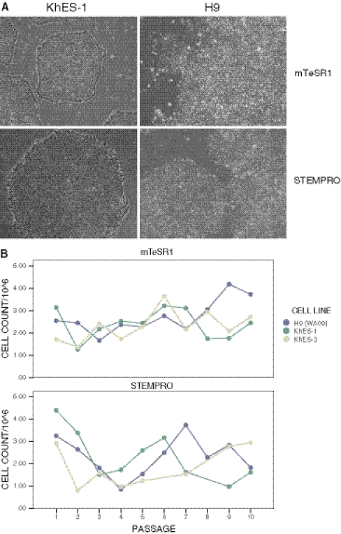
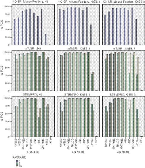
There are many reports of defined culture systems for the propagation of human embryonic stem cells in the absence of feeder cell support, but no previous study has undertaken a multi-laboratory comparison of these diverse methodologies. In this study, five separate laboratories, each with experience in human embryonic stem cell culture, used a panel of ten embryonic stem cell lines (including WA09 as an index cell line common to all laboratories) to assess eight cell culture methods, with propagation in the presence of Knockout Serum Replacer, FGF-2, and mouse embryonic fibroblast feeder cell layers serving as a positive control. The cultures were assessed for up to ten passages for attachment, death, and differentiated morphology by phase contrast microscopy, for growth by serial cell counts, and for maintenance of stem cell surface marker expression by flow cytometry. Of the eight culture systems, only the control and those based on two commercial media, mTeSR1 and STEMPRO, supported maintenance of most cell lines for ten passages. Cultures grown in the remaining media failed before this point due to lack of attachment, cell death, or overt cell differentiation. Possible explanations for relative success of the commercial formulations in this study, and the lack of success with other formulations from academic groups compared to previously published results, include: the complex combination of growth factors present in the commercial preparations; improved development, manufacture, and quality control in the commercial products; differences in epigenetic adaptation to culture in vitro between different ES cell lines grown in different laboratories.


Figures

Figure 1.
Figure 1.
(A) Summary test results. (B) Results of the retest of the media by UKSCB. For most tests new growth factors were obtained however for testing of medium no. 3 three different batches of Activin A were used. ISCI-GF: Original growth factor batch used in the ISCI study. UKSCB-GF: New Growth factor obtained for the retest. LV-GF: Activin A obtained from the originating laboratory.
Figure 1.
Figure 1.
(A) Summary test results. (B) Results of the retest of the media by UKSCB. For most tests new growth factors were obtained however for testing of medium no. 3 three different batches of Activin A were used. ISCI-GF: Original growth factor batch used in the ISCI study. UKSCB-GF: New Growth factor obtained for the retest. LV-GF: Activin A obtained from the originating laboratory.
Figure 2.
Figure 2.
Representative photomicrographs and cell counts. (A) Photomicrographs of KhES-1 and H9 (WA09) respectively grow to ten passages in mTeSR1 and STEMPRO, respectively. (B) Representative cell counts from each passage for the cell lines WA09 (H9), KhES-1, and KhES-3.
Figure 3.
Figure 3.
Representative flow cytometry data. Representative flow cytometry data expressed as percentage of cells called positive for three cell lines H9, KhES-1, and KhES-3 at passage 0 in Knockout Serum Replacer, FGF-2, and mouse embryonic fibroblast feeder cells and at passage 5 and 10 for cells grown in mTeSR1 and STEMPRO.

Similar articles

Cited by

References

    1. Adewumi O, Aflatoonian B, Ahrlund-Richter L, Amit M, Andrews PW, Beighton G, Bello PA, Benvenisty N, Berry LS, Bevan S, et al. Characterization of human embryonic stem cell lines by the International Stem Cell Initiative. Nat. Biotechnol. 2007;25:803–816. doi: 10.1038/nbt1318. - DOI - PubMed
    1. Amit M, Itskovitz-Eldor J. Maintenance of human embryonic stem cells in animal serum- and feeder layer-free culture conditions. Methods Mol. Biol. 2006;331:105–113. - PubMed
    1. Andrews PW, Benvenisty N, McKay R, Pera MF, Rossant J, Semb H, Stacey GN. The International Stem Cell Initiative: toward benchmarks for human embryonic stem cell research. Nat. Biotechnol. 2005;23:795–797. doi: 10.1038/nbt0705-795. - DOI - PubMed
    1. Andrews PW, Goodfellow PN, Shevinsky LH, Bronson DL, Knowles BB. Cell-surface antigens of a clonal human embryonal carcinoma cell line: morphological and antigenic differentiation in culture. Int. J. Cancer. 1982;29:523–531. doi: 10.1002/ijc.2910290507. - DOI - PubMed
    1. Beattie GM, Lopez AD, Bucay N, Hinton A, Firpo MT, King CC, Hayek A. Activin A maintains pluripotency of human embryonic stem cells in the absence of feeder layers. Stem Cells. 2005;23:489–495. doi: 10.1634/stemcells.2004-0279. - DOI - PubMed

Publication types

LinkOut - more resources