HL-1 cells express an inwardly rectifying K+ current activated via muscarinic receptors comparable to that in mouse atrial myocytes
- PMID: 20186548
- PMCID: PMC2872014
- DOI: 10.1007/s00424-010-0799-z
HL-1 cells express an inwardly rectifying K+ current activated via muscarinic receptors comparable to that in mouse atrial myocytes
Abstract
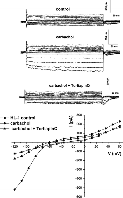
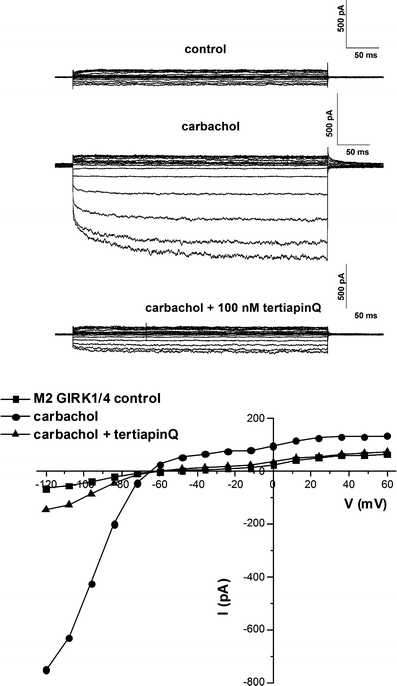
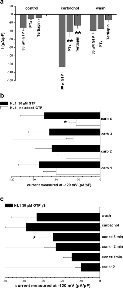
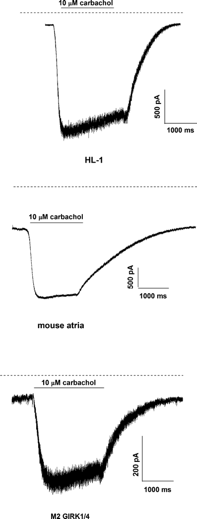
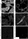
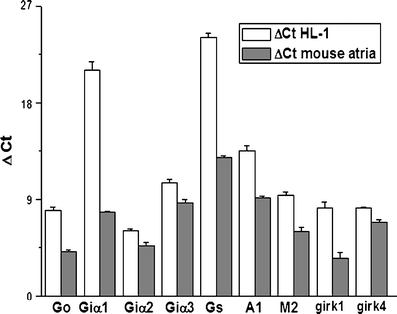
An inwardly rectifying K(+) current is present in atrial cardiac myocytes that is activated by acetylcholine (I(KACh)). Physiologically, activation of the current in the SA node is important in slowing the heart rate with increased parasympathetic tone. It is a paradigm for the direct regulation of signaling effectors by the Gbetagamma G-protein subunit. Many questions have been addressed in heterologous expression systems with less focus on the behaviour in native myocytes partly because of the technical difficulties in undertaking comparable studies in native cells. In this study, we characterise a potassium current in the atrial-derived cell line HL-1. Using an electrophysiological approach, we compare the characteristics of the potassium current with those in native atrial cells and in a HEK cell line expressing the cloned Kir3.1/3.4 channel. The potassium current recorded in HL-1 is inwardly rectifying and activated by the muscarinic agonist carbachol. Carbachol-activated currents were inhibited by pertussis toxin and tertiapin-Q. The basal current was time-dependently increased when GTP was substituted in the patch-clamp pipette by the non-hydrolysable analogue GTPgammaS. We compared the kinetics of current modulation in HL-1 with those of freshly isolated atrial mouse cardiomyocytes. The current activation and deactivation kinetics in HL-1 cells are comparable to those measured in atrial cardiomyocytes. Using immunofluorescence, we found GIRK4 at the membrane in HL-1 cells. Real-time RT-PCR confirms the presence of mRNA for the main G-protein subunits, as well as for M2 muscarinic and A1 adenosine receptors. The data suggest HL-1 cells are a good model to study IKAch.
Figures
Similar articles
-
Differential effects of genetically-encoded Gβγ scavengers on receptor-activated and basal Kir3.1/Kir3.4 channel current in rat atrial myocytes.Cell Signal. 2014 Jun;26(6):1182-92. doi: 10.1016/j.cellsig.2014.02.007. Epub 2014 Feb 24. Cell Signal. 2014. PMID: 24576551
-
Differential subunit composition of the G protein-activated inward-rectifier potassium channel during cardiac development.J Clin Invest. 2004 Oct;114(7):994-1001. doi: 10.1172/JCI15925. J Clin Invest. 2004. PMID: 15467839 Free PMC article.
-
A new potassium ion current induced by stimulation of M2 cholinoreceptors in fish atrial myocytes.J Exp Biol. 2014 May 15;217(Pt 10):1745-51. doi: 10.1242/jeb.098509. Epub 2014 Feb 13. J Exp Biol. 2014. PMID: 24526726
-
Pharmacological Conversion of a Cardiac Inward Rectifier into an Outward Rectifier Potassium Channel.Mol Pharmacol. 2016 Sep;90(3):334-40. doi: 10.1124/mol.116.104950. Epub 2016 May 31. Mol Pharmacol. 2016. PMID: 27247338 Review.
-
The Role of KACh Channels in Atrial Fibrillation.Cells. 2024 Jun 10;13(12):1014. doi: 10.3390/cells13121014. Cells. 2024. PMID: 38920645 Free PMC article. Review.
Cited by
-
Subtype-dependent regulation of Gβγ signalling.Cell Signal. 2021 Jun;82:109947. doi: 10.1016/j.cellsig.2021.109947. Epub 2021 Feb 11. Cell Signal. 2021. PMID: 33582184 Free PMC article. Review.
-
Higher-order transient membrane protein structures.Proc Natl Acad Sci U S A. 2025 Jan 7;122(1):e2421275121. doi: 10.1073/pnas.2421275121. Epub 2024 Dec 31. Proc Natl Acad Sci U S A. 2025. PMID: 39739811 Free PMC article.
-
Lipopolysaccharide prolongs action potential duration in HL-1 mouse cardiomyocytes.Am J Physiol Cell Physiol. 2012 Oct 15;303(8):C825-33. doi: 10.1152/ajpcell.00173.2012. Epub 2012 Aug 15. Am J Physiol Cell Physiol. 2012. PMID: 22895260 Free PMC article.
-
Critical evaluation of KCNJ3 gene product detection in human breast cancer: mRNA in situ hybridisation is superior to immunohistochemistry.J Clin Pathol. 2016 Dec;69(12):1116-1121. doi: 10.1136/jclinpath-2016-203798. Epub 2016 Oct 3. J Clin Pathol. 2016. PMID: 27698251 Free PMC article.
-
Do caveolae have a role in the fidelity and dynamics of receptor activation of G-protein-gated inwardly rectifying potassium channels?J Biol Chem. 2010 Sep 3;285(36):27817-26. doi: 10.1074/jbc.M110.103598. Epub 2010 Jun 18. J Biol Chem. 2010. PMID: 20562107 Free PMC article.
References
-
- Wellner-Kienitz MC, Bender K, Meyer T, Bunemann M, Pott L. Overexpressed A(1) adenosine receptors reduce activation of acetylcholine-sensitive K(+) current by native muscarinic M(2) receptors in rat atrial myocytes. Circ Res. 2000;86:643–648. - PubMed
Publication types
MeSH terms
Substances
Grants and funding
LinkOut - more resources
Full Text Sources
Other Literature Sources
Medical
Miscellaneous

