

An improved collagen scaffold for skeletal regeneration
- PMID: 20186736
- PMCID: PMC2891373
- DOI: 10.1002/jbm.a.32694
An improved collagen scaffold for skeletal regeneration
Abstract
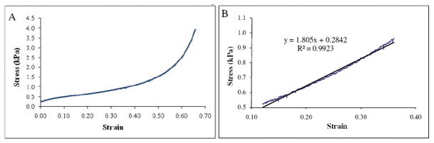
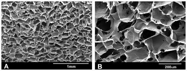
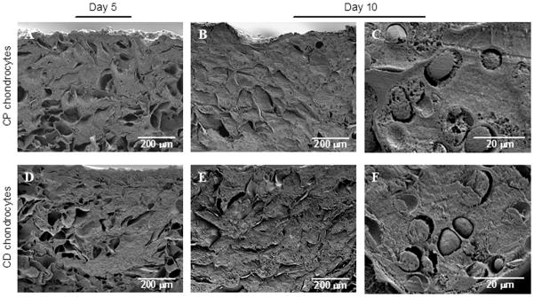
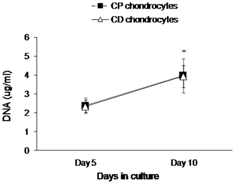
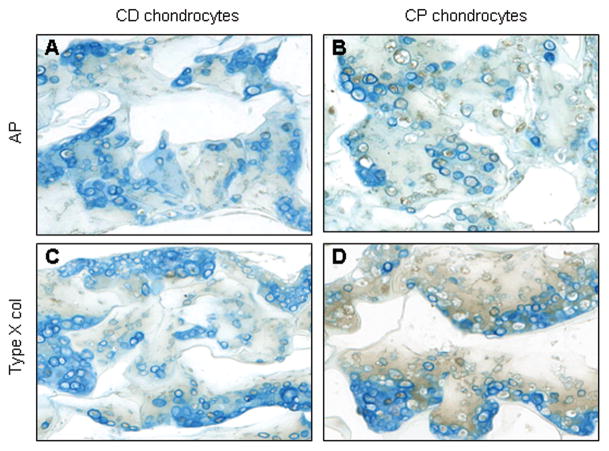
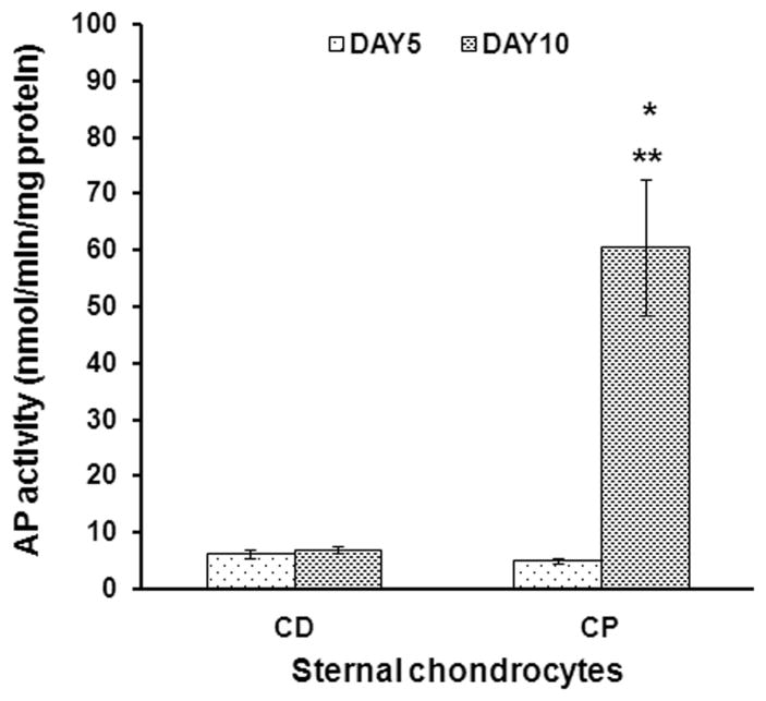
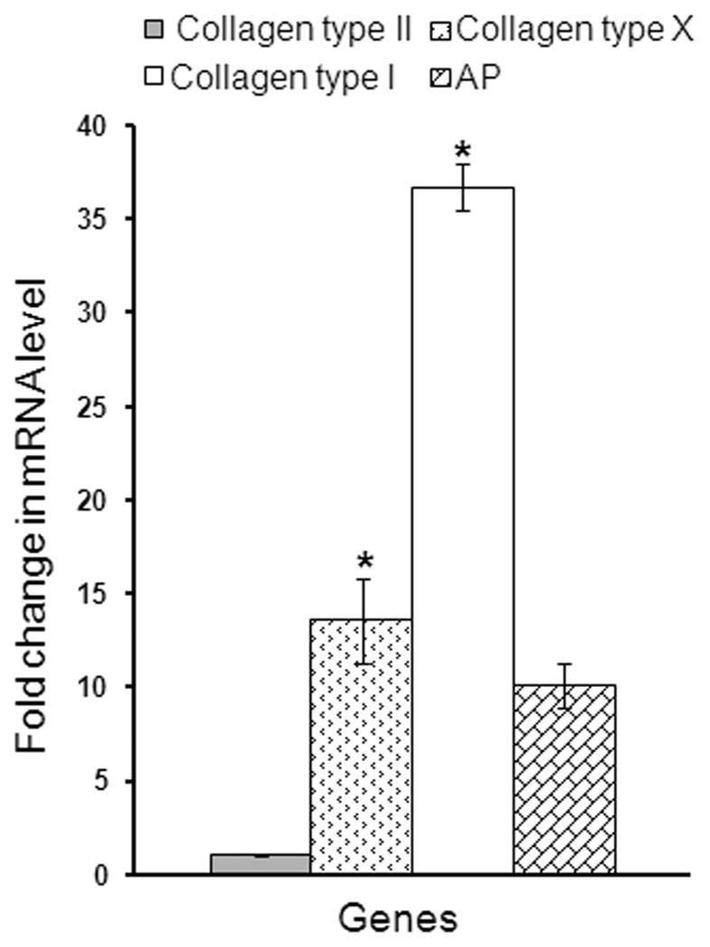
Bone repair and regeneration is one of the most extensively studied areas in the field of tissue engineering. All of the current tissue engineering approaches to create bone focus on intramembranous ossification, ignoring the other mechanism of bone formation, endochondral ossification. We propose to create a transient cartilage template in vitro, which could serve as an intermediate for bone formation by the endochondral mechanism once implanted in vivo. The goals of the study are (1) to prepare and characterize type I collagen sponges as a scaffold for the cartilage template, and (2) to establish a method of culturing chondrocytes in type I collagen sponges and induce cell maturation. Collagen sponges were generated from a 1% solution of type I collagen using a freeze/dry technique followed by UV light crosslinking. Chondrocytes isolated from two locations in chick embryo sterna were cultured in these sponges and treated with retinoic acid to induce chondrocyte maturation and extracellular matrix deposition. Material strength testing as well as microscopic and biochemical analyzes were conducted to evaluate the properties of sponges and cell behavior during the culture period. We found that our collagen sponges presented improved stiffness and supported chondrocyte attachment and proliferation. Cells underwent maturation, depositing an abundant extracellular matrix throughout the scaffold, expressing high levels of type X collagen, type I collagen and alkaline phosphatase. These results demonstrate that we have created a transient cartilage template with potential to direct endochondral bone formation after implantation.
(c) 2010 Wiley Periodicals, Inc.
Figures
References
-
- Pountos I, Jones E, Tzioupis C, McGonagle D, Giannoudis PV. Growing bone and cartilage. The role of mesenchymal stem cells. J Bone Joint Surg Br. 2006;88(4):421–6. - PubMed
-
- Newman AP. Articular cartilage repair. Am J Sports Med. 1998;26(2):309–24. - PubMed
-
- Tomihisa K, Tomoo M, Toshitaka T, Tomoyuki S. New bone formation around porous hydroxyapatite wedge implanted in opening wedge high tibial osteotomy in patients with osteoarthritis. Biomaterials. 2001;22(12):1579–1582. - PubMed
-
- Weiss P, Layrolle P, Clergeau LP, Enckel B, Pilet P, Amouriq Y, Daculsi G, Giumelli B. The safety and efficacy of an injectable bone substitute in dental sockets demonstrated in a human clinical trial. Biomaterials. 2007;28(22):3295–3305. - PubMed
-
- Verlaan JJ, Oner FC, Dhert WJ. Anterior spinal column augmentation with injectable bone cements. Biomaterials. 2006;27(3):290–301. - PubMed
Publication types
MeSH terms
Substances
Grants and funding
LinkOut - more resources
Full Text Sources
Other Literature Sources
