. 2010 Mar;62(3):791-801.
doi: 10.1002/art.27305.

Autophagy is a protective mechanism in normal cartilage, and its aging-related loss is linked with cell death and osteoarthritis

Affiliations

Autophagy is a protective mechanism in normal cartilage, and its aging-related loss is linked with cell death and osteoarthritis

Beatriz Caramés et al. Arthritis Rheum. 2010 Mar.

Abstract

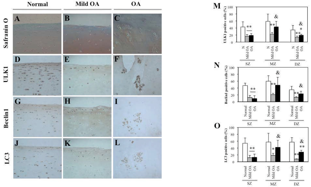
Objective: Autophagy is a process for turnover of intracellular organelles and molecules that protects cells during stress responses. We undertook this study to evaluate the potential roles of Unc-51-like kinase 1 (ULK1), an inducer of autophagy, Beclin1, a regulator of autophagy, and microtubule-associated protein 1 light chain 3 (LC3), which executes autophagy, in the development of osteoarthritis (OA) and in cartilage cell death.

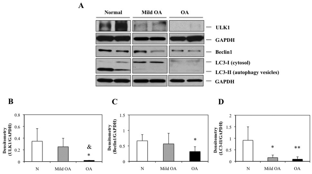
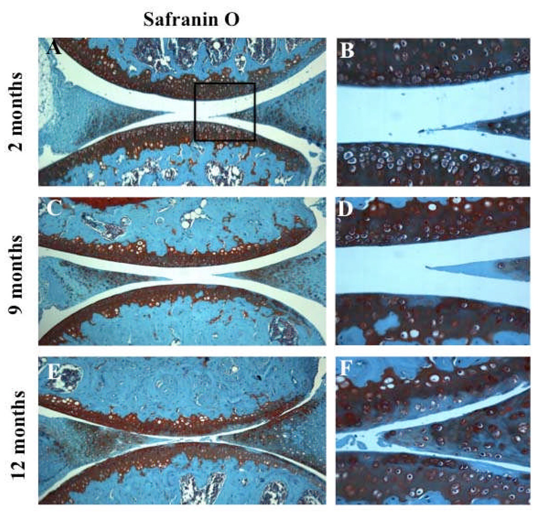
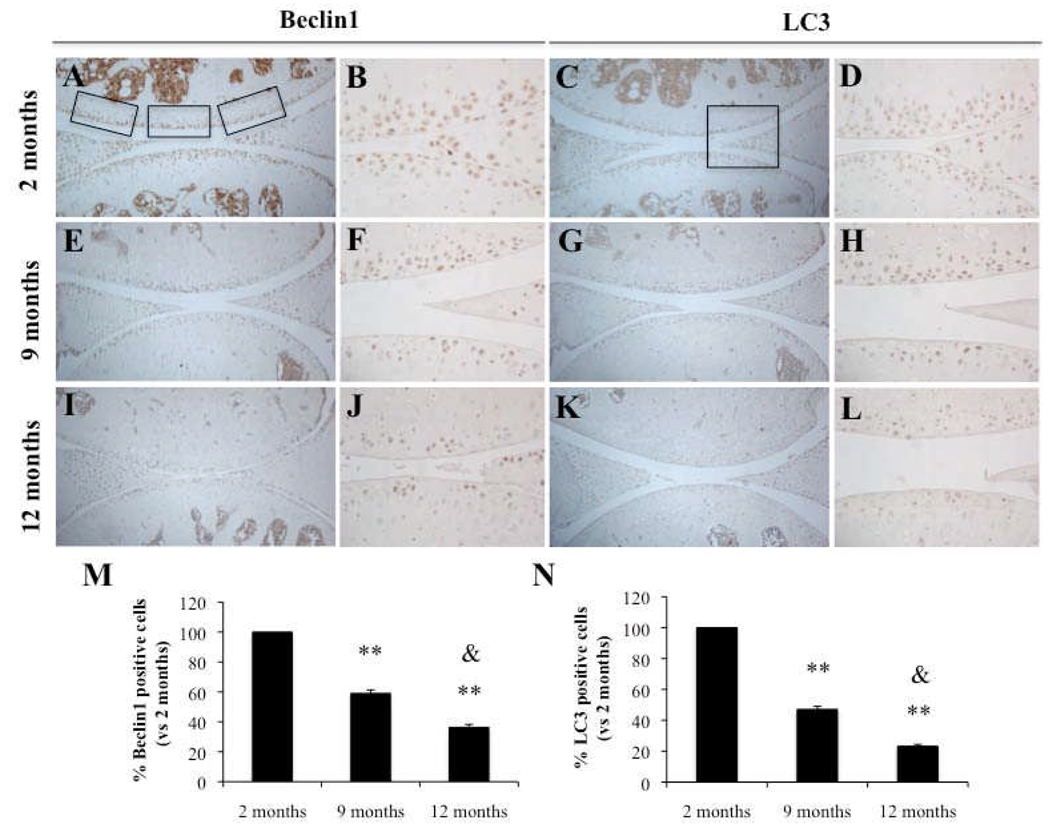
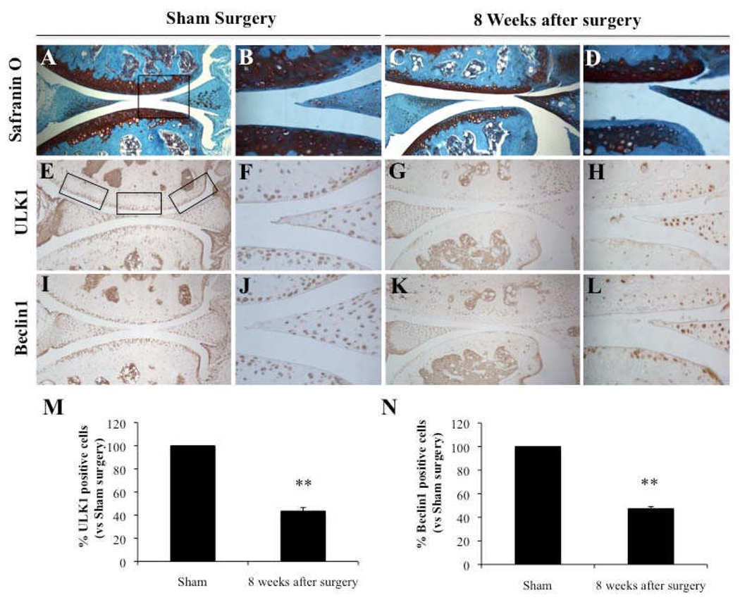
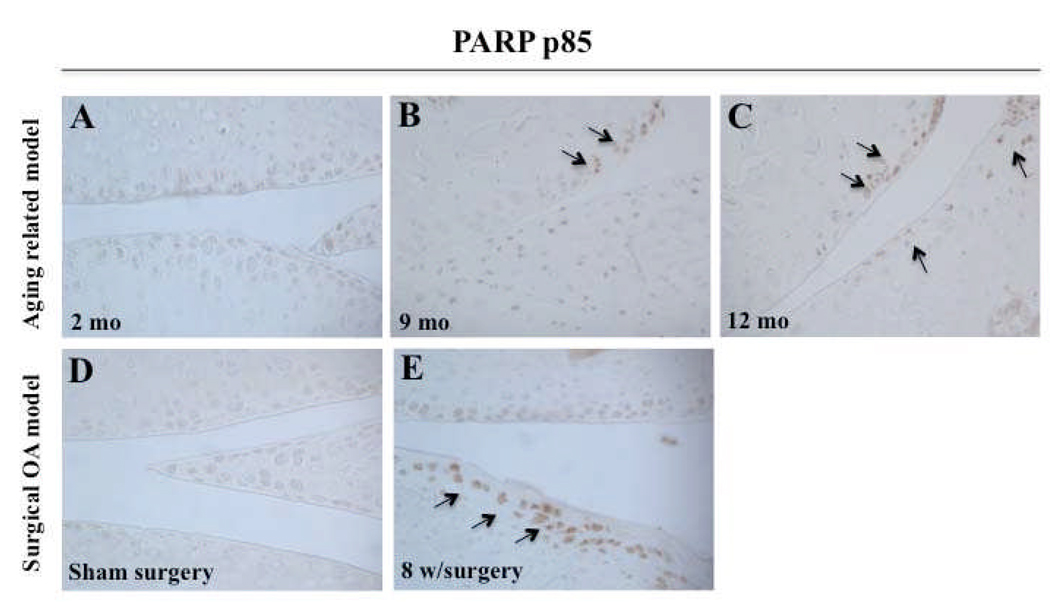
Methods: Expression of ULK1, Beclin1, and LC3 was analyzed in normal and OA human articular cartilage and in knee joints of mice with aging-related and surgically induced OA, using immunohistochemistry and Western blotting. Poly(ADP-ribose) polymerase (PARP) p85 expression was used to determine the correlation between cell death and autophagy.

Results: ULK1, Beclin1, and LC3 were constitutively expressed in normal human articular cartilage. ULK1, Beclin1, and LC3 protein expression was reduced in OA chondrocytes and cartilage, but these 3 proteins were strongly expressed in the OA cell clusters. In mouse knee joints, loss of glycosaminoglycans (GAGs) was observed at ages 9 months and 12 months and in the surgical OA model, 8 weeks after knee destabilization. Expression of ULK1, Beclin1, and LC3 decreased together with GAG loss, while PARP p85 expression was increased.

Conclusion: Autophagy may be a protective or homeostatic mechanism in normal cartilage. In contrast, human OA and aging-related and surgically induced OA in mice are associated with a reduction and loss of ULK1, Beclin1, and LC3 expression and a related increase in apoptosis. These results suggest that compromised autophagy represents a novel mechanism in the development of OA.


Figures

Figure 1
Figure 1
ULK1, Beclin1 and LC3 expression is reduced in human osteoarthritic (OA) cartilage. A–C, Representative sections of 6 normal (N) cartilage, 4 mild OA and 6 OA cartilage samples as shown by Safranin O. D–F, G–I, J–L, Immunohistochemical analysis with anti-ULK1, Beclin1 and LC3, respectively. M–O, Quantification of ULK1, Beclin1 and LC3-positive cells in N, mild OA and OA cartilage. In mild OA, the percentage of ULK1, Beclin1 and LC3-positive cells was significantly reduced in superficial (SZ), middle (MZ) and deep zone (DZ) compared to N cartilage. Values are the mean ± SD. ** = P < 0.01 versus N cartilage; * = P < 0.05 versus N cartilage. In OA cartilage, ULK1, Beclin1 and LC3 expression were significantly reduced in SZ compared to N cartilage. In DZ only ULK1 and LC3 expression was significantly reduced. Values are the mean ± SD. ** = P < 0.01 versus N cartilage; * = P < 0.05 versus N cartilage in DZ. Furthermore, ULK1, Beclin1 and LC3 expression was significantly increased in OA cartilage compared to mild OA in MZ and DZ. Values are the mean ± SD. & = P < 0.05 versus Mild OA. Magnification: A–L x40.
Figure 2
Figure 2
ULK1, Beclin1 and LC3 expression is decreased in human osteoarthritic (OA) chondrocytes. A, Total protein from N, mild OA and OA chondrocytes was analyzed by Western blotting using anti-ULK1, Beclin1, LC3 and GAPDH as described in methods. The images shown are representative of 4 N, mild OA and OA donors for ULK1 and 6 N, 4 mild OA and 6 OA donors for Beclin1 and LC3. Representative blots are shown along with the numeric data obtained by densitometry. B–D, Densitometry analysis showed a significant decreased of protein expression by 17-fold, 2.1-fold and 9-fold for ULK1, Beclin1 and LC3-II, respectively, in OA chondrocytes compared to N chondrocytes. Values are the mean ± SD. * = P < 0.05 versus N cartilage and ** = P < 0.01 versus N cartilage. On the other hand, ULK1 protein expression was significantly reduced in OA chondrocytes compared to mild OA. Values are the mean ± SD. & = P < 0.05 versus mild OA.
Figure 3
Figure 3
Aging-related loss of glycosaminoglycans (GAGs) in mouse joints. A–F, Knee joints from 2 months (n=4), 9 months (n=4) and 12 months (n=4) old C57Bl/6J mice were analyzed by Safranin O staining. Magnification: A,C,E x10; B,D,F x40.
Figure 4
Figure 4
Aging-related reduction in Beclin1 and LC3 expression in mouse joints. Knee joints from 2, 9 and 12 month old C57Bl/6J mice were analyzed by immunohistochemistry (IHC) for Beclin1 (A–B, E–F, I–J) and LC3 (C–D, G–H, K–L). M–N, Quantification of Beclin1 and LC3-positive cells at 2, 9 and 12 months. Beclin1 and LC3-positive cells were significantly decreased at 9 and 12 months compared to 2 months. Values are the mean ± SD. ** = P < 0.01 versus 2 months old. Furthermore, Beclin1 and LC3-positive cells were significantly reduced at 12 months compared to 9 months. Values are the mean ± SD. & = P < 0.05 versus 9 months old. Knee joints from C57BL/6JWT mice age 2 months (n=4), 9 months (n=4) and 12 months (n=4) were examined. Magnification: A,C,E,G,I,K x10; B,D,F,H,J,L x40.
Figure 5
Figure 5
Reduction in ULK1 and Beclin1 expression in knee joints from mice with surgical OA. Knee joints from 2 months old C57Bl/6J mice with Sham surgery (n=4) and with surgical OA induced by transection of the medial meniscotibial ligament and the medial collateral ligament (MMTL+MCL) at 8 weeks (n=4) were analyzed by Safranin O staining (A–D) or for ULK1 (E–H) and Beclin1 (I–L). M–N, Quantification of ULK1 and Beclin1-positive cells. The expression was significantly decreased at 8 weeks after surgery. Values are the mean ± SD. ** = P < 0.01 versus Sham surgery. Magnification: A,C,E,G,I,K x10; B,D,F,H,J,L x40.
Figure 6
Figure 6
Increased cell death in aging-related and surgical OA in mouse joints. A–C, Knee joints from 2, 9, 12 month old C57Bl/6J mice were used to evaluate PARP p85 expression by immunohistochemistry (IHC). Knee joints from C57BL/6JWT mice aged 2 months (n=3), 9 months (n=3) and 12 months (n=3) were examined. D–E, Knee joints from 2 months old C57Bl/6J mice with sham surgery (n=3) and surgical OA at 8 weeks (n=3) were evaluated for PARP p85 expression by IHC. Magnification: x40.

References

    1. Ruiz-Romero C, Calamia V, Mateos J, Carreira V, Martínez-Gomariz M, Fernández M, et al. Mitochondrial dysregulation of osteoarthritic human articular chondrocytes analyzed by proteomics: A decrease in mitochondrial superoxide dismutase points to a redox imbalance. Mol Cell Proteomics. 2009;8:172–189. - PMC - PubMed
    1. Taniguchi N, Caramés B, Ronfani L, Ulmer U, Komiya S, Bianchi ME, et al. Aging-related loss of the chromatin protein HMGB2 in articular cartilage is linked to reduced cellularity and osteoarthritis. Proc Natl Acad Sci U S A. 2009;27(106):1181–1186. - PMC - PubMed
    1. Pennock AT, Robertson CM, Emmerson BC, Harwood FL, Amiel D. Role of apoptotic and matrix-degrading genes in articular cartilage and meniscus of mature and aged rabbits during development of osteoarthritis. Arthritis Rhem. 2007;56:1529–1536. - PubMed
    1. Aigner T, Haag J, Martin J, Buckwalter J. Osteoarthritis: aging of matrix and cells--going for a remedy. Curr Drug Targets. 2007;8:325–331. - PubMed
    1. Vellai T. Autophagy genes and ageing. Autophagy genes and ageing. Cell Death Differ. 2009;16:94–102. - PubMed

Publication types

MeSH terms