. 2010 Mar;63(3):736-44.
doi: 10.1002/mrm.22227.

Effect of blood flow on double inversion recovery vessel wall MRI of the peripheral arteries: quantitation with T2 mapping and comparison with flow-insensitive T2-prepared inversion recovery imaging

Affiliations

Effect of blood flow on double inversion recovery vessel wall MRI of the peripheral arteries: quantitation with T2 mapping and comparison with flow-insensitive T2-prepared inversion recovery imaging

Ryan Brown et al. Magn Reson Med. 2010 Mar.

Erratum in

  • Magn Reson Med. 2010 Nov;64(5):1540

Abstract

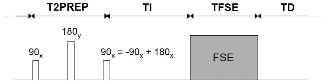
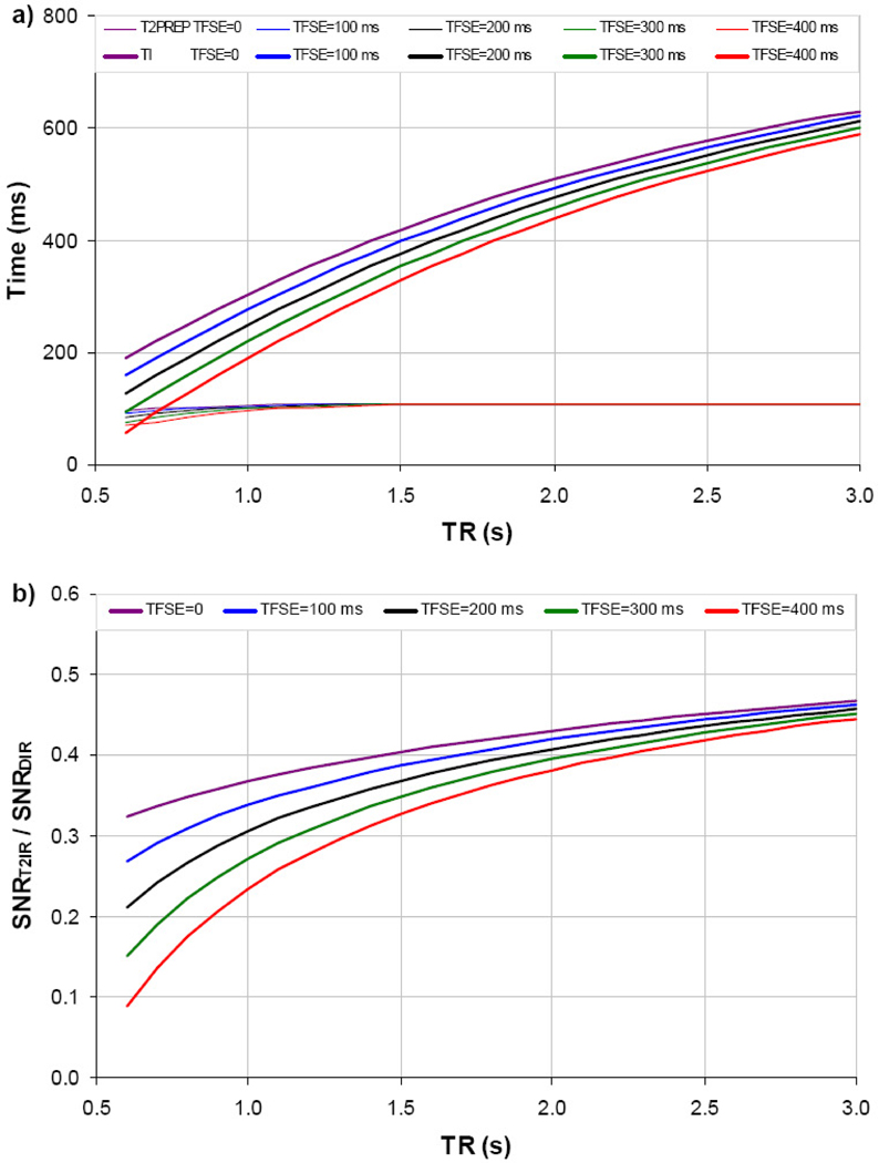
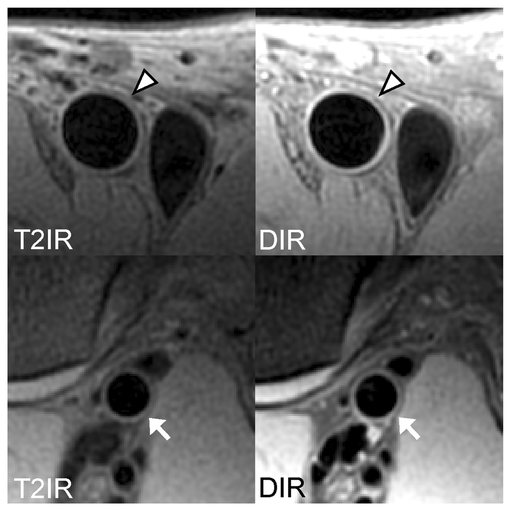
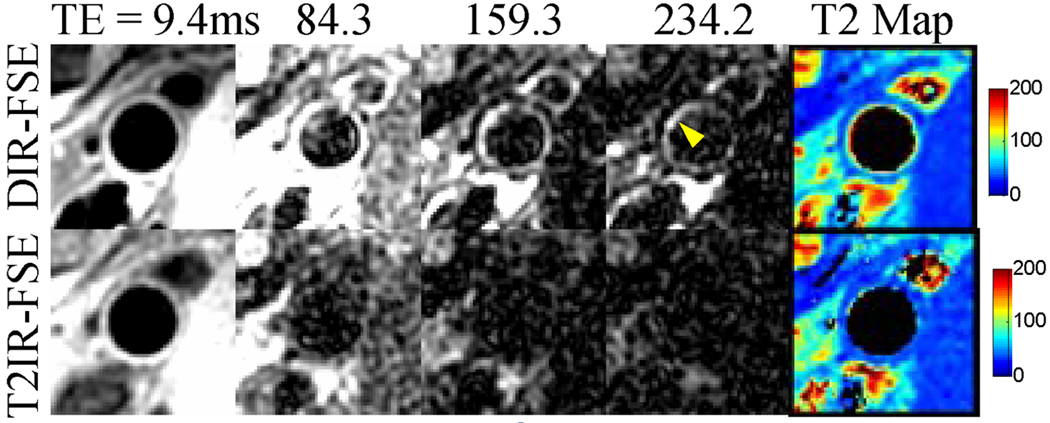
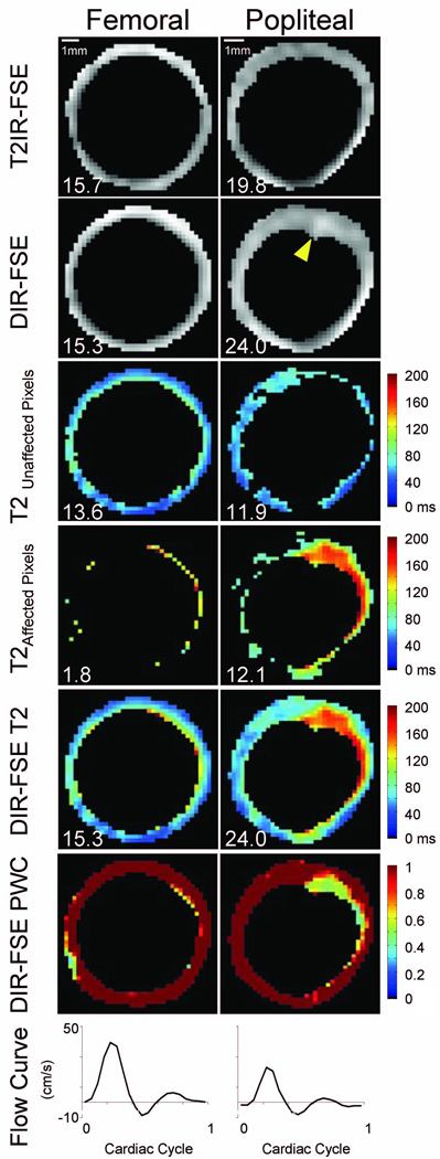
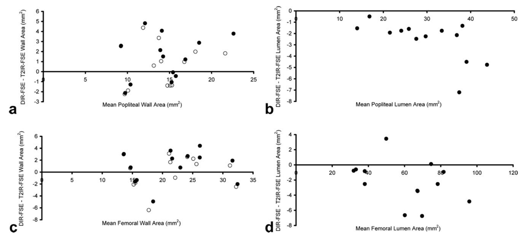
Blood suppression in the lower extremities using flow-reliant methods such as double inversion recovery may be problematic due to slow blood flow. T(2) mapping using fast spin echo (FSE) acquisition was utilized to quantitate the effectiveness of double inversion recovery blood suppression in 13 subjects and showed that 25 +/- 12% of perceived vessel wall pixels in the popliteal arteries contained artifactual blood signal. To overcome this problem, a flow-insensitive T(2)-prepared inversion recovery sequence was implemented and optimal timing parameters were calculated for FSE acquisition. Black blood vessel wall imaging of the popliteal and femoral arteries was performed using two-dimensional T(2)-prepared inversion recovery-FSE in the same 13 subjects. Comparison with two-dimensional double inversion recovery-FSE showed that T(2)-prepared inversion recovery-FSE reduced wall-mimicking blood artifacts that inflated double inversion recovery-FSE vessel wall area measurements in the popliteal artery.


Figures

Figure 1
Figure 1
Schematic of the implemented T2IR-FSE sequence for flow-insensitive BB imaging of the vessel wall. The T2IR magnetization preparation combines two commonly used contrast-enhancing techniques: T2 preparation (consisting of 90x tip-down, 180y refocusing, and −90x tip-up RF pulses) for T2 contrast, and inversion recovery (consisting of a 180x inversion pulse) for T1 contrast. Note that the −90x tip-up of the T2 preparation and the 180x inversion pulse can be conveniently combined into a single 90x pulse as shown. Given repetition time TR = T2PREP + TI + TFSE + TD, the timing parameters (T2PREP, TI and TD) are chosen to null the blood signal while maximizing the wall signal at the beginning of the FSE acquisition (see text for details). Note that all RF pulses are spatially non-selective and correspondingly the T2IR technique can provide global suppression of blood regardless of its movement, a major advantage over the conventional DIR technique.
Figure 2
Figure 2
Optimal T2PREP and TI as a function of TR and TFSE for BB T2IR-FSE vessel wall imaging at 1.5 T (a). Corresponding theoretical relative wall SNR between optimized T2IR-FSE and DIR-FSE is shown in b.
Figure 3
Figure 3
Concordant femoral (arrowheads) and popliteal (arrows) vessel wall images obtained with T2IR-FSE and DIR-FSE. Note the effective arterial blood suppression by both techniques.
Figure 4
Figure 4
Blood artifact (arrowhead) at the popliteal wall-lumen border in the DIR-FSE images (top row). While this artifact is not obvious with effective TE of 9.4 ms, it is conspicuous in images acquired at longer effective TEs where wall components and surrounding muscles with short T2s have decayed. The DIR-FSE T2 map shows elevated T2 values along the wall-lumen interface, indicating the presence of blood signal. These artifacts are absent in the T2IR-FSE images and corresponding T2 map (bottom row).
Figure 5
Figure 5
Representative data set highlighting DIR-FSE popliteal blood artifact. Top row: segmented T2IR-FSE images at effective TE = 9.4 ms; second row: segmented DIR-FSE images at effective TE = 9.4 ms; third row: DIR-FSE T2 maps for unaffected vessel wall pixels; fourth row: DIR-FSE T2 maps for pixels affected by slow flowing blood; fifth row: collective DIR-FSE T2 maps; sixth row: DIR-FSE partial wall content (PWC) maps; and bottom row: blood velocity curves. The arrowhead in the DIR-FSE popliteal image indicates an area of incomplete blood suppression. The scale is indicated in the top row. Corresponding areas (in mm2) are inset for reference.
Figure 6
Figure 6
Bland-Altman plots of wall area (a, b) and lumen area (c, d) measurements obtained with 2D DIR-FSE and T2IR-FSE imaging. Before DIR-FSE partial wall content correction (solid circles), agreement was 1.4 ± 2.2 mm2 for the popliteal wall (a) and 0.9 ± 2.7 mm2 for the femoral wall (b). After DIR-FSE partial wall content correction (open circles), agreement was 0.6 ± 2.1 mm2 for the popliteal wall and 0.2 ± 2.8 mm2 for the femoral wall. Popliteal lumen area bias was −2.6 ± 1.8 mm2 (c) while femoral lumen area bias was −2.3 ± 2.8 mm2 (d).

Similar articles

Cited by

References

    1. Malayeri AA, Natori S, Bahrami H, Bertoni AG, Kronmal R, Lima JA, Bluemke DA. Relation of aortic wall thickness and distensibility to cardiovascular risk factors (from the Multi-Ethnic Study of Atherosclerosis [MESA]) Am J Cardiol. 2008;102(4):491–496. - PMC - PubMed
    1. Amarenco P, Cohen A, Tzourio C, Bertrand B, Hommel M, Besson G, Chauvel C, Touboul PJ, Bousser MG. Atherosclerotic disease of the aortic arch and the risk of ischemic stroke. N Engl J Med. 1994;331(22):1474–1479. - PubMed
    1. Smilde TJ, van den Berkmortel FW, Boers GH, Wollersheim H, de Boo T, van Langen H, Stalenhoef AF. Carotid and femoral artery wall thickness and stiffness in patients at risk for cardiovascular disease, with special emphasis on hyperhomocysteinemia. Arterioscler Thromb Vasc Biol. 1998;18(12):1958–1963. - PubMed
    1. Demarco JK, Rutt BK, Clarke SE. Carotid plaque characterization by magnetic resonance imaging: review of the literature. Top Magn Reson Imaging. 2001;12(3):205–217. - PubMed
    1. Underhill HR, Yuan C, Zhao XQ, Kraiss LW, Parker DL, Saam T, Chu B, Takaya N, Liu F, Polissar NL, Neradilek B, Raichlen JS, Cain VA, Waterton JC, Hamar W, Hatsukami TS. Effect of rosuvastatin therapy on carotid plaque morphology and composition in moderately hypercholesterolemic patients: a high-resolution magnetic resonance imaging trial. Am Heart J. 2008;155(3):584, e581–e588. - PubMed

Publication types