. 2010 Apr 1;201(7):993-9.
doi: 10.1086/651132.

Severity of pneumonia due to new H1N1 influenza virus in ferrets is intermediate between that due to seasonal H1N1 virus and highly pathogenic avian influenza H5N1 virus

Affiliations

Severity of pneumonia due to new H1N1 influenza virus in ferrets is intermediate between that due to seasonal H1N1 virus and highly pathogenic avian influenza H5N1 virus

Judith M A van den Brand et al. J Infect Dis. .

Abstract

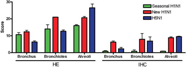
Background: The newly emerged influenza A(H1N1) virus (new H1N1 virus) is causing the first influenza pandemic of this century. Three influenza pandemics of the previous century caused variable mortality, which largely depended on the development of severe pneumonia. However, the ability of the new H1N1 virus to cause pneumonia is poorly understood.

Methods: The new H1N1 virus was inoculated intratracheally into ferrets. Its ability to cause pneumonia was compared with that of seasonal influenza H1N1 virus and highly pathogenic avian influenza (HPAI) H5N1 virus by using clinical, virological, and pathological analyses.

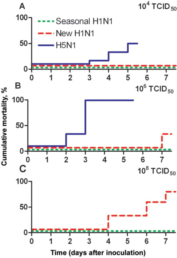
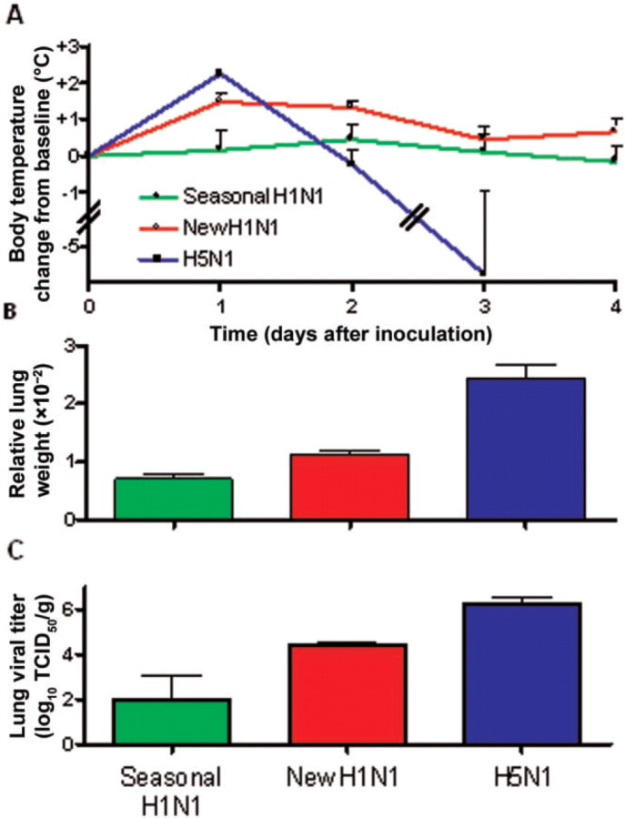
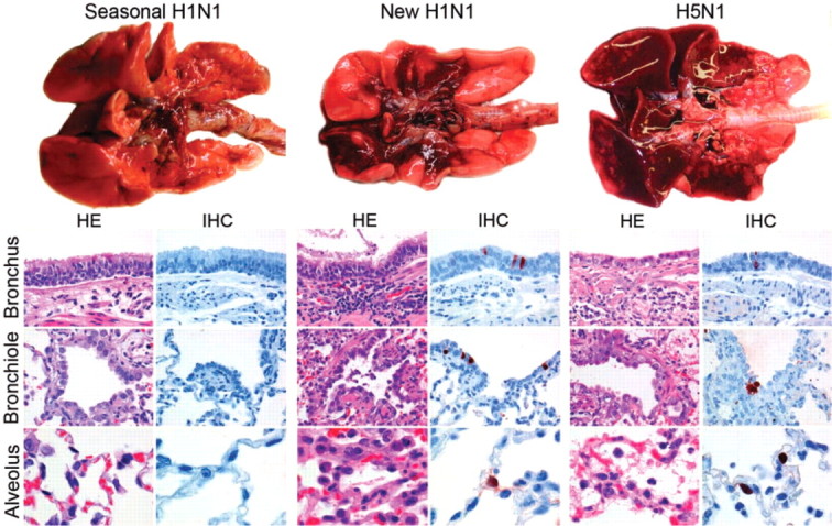
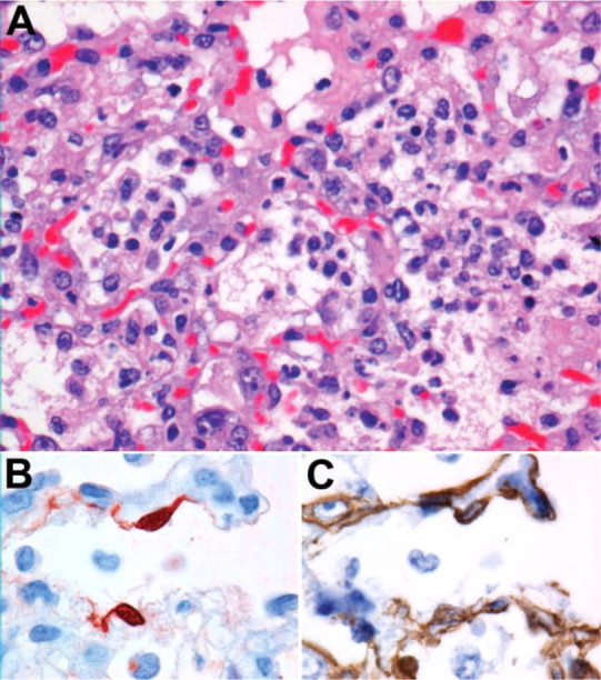
Results: Our results showed that the new H1N1 virus causes pneumonia in ferrets intermediate in severity between that caused by seasonal H1N1 virus and by HPAI H5N1 virus. The new H1N1 virus replicated well throughout the lower respiratory tract and more extensively than did both seasonal H1N1 virus (which replicated mainly in the bronchi) and HPAI H5N1 virus (which replicated mainly in the alveoli). High loads of new H1N1 virus in lung tissue were associated with diffuse alveolar damage and mortality.

Conclusions: The new H1N1 virus may be intrinsically more pathogenic for humans than is seasonal H1N1 virus.


Figures

Figure 1.
Figure 1.
Cumulative mortality rates of ferrets inoculated with different influenza viruses. Ferrets were intratracheally inoculated with seasonal H1N1 (n = 3), new H1N1 (n = 6), or highly pathogenic avian influenza (HPAI) H5N1 (n = 6) influenza viruses at a dose of 104 (A), 106 (B), or 108 (C) median tissue culture infective dose (TCID50). Cumulative mortality for new H1N1 virus was intermediate between that for seasonal H1N1 virus and that for HPAI H5N1 virus.
Figure 2.
Figure 2.
Body temperatures, relative lung weights and lung viral titers of ferrets inoculated with different influenza viruses. The ferrets were intratracheally inoculated with seasonal H1N1 (n = 3), new H1N1 (n = 6) or highly pathogenic avian influenza (HPAI) H5N1 (n = 6)influenza viruses at a dose of 106 median tissue culture infective dose (TCID50). The increase in body temperature (A), the relative lung weight (B), and the lung viral titer (C) of the new H1N1 virus group were intermediate between those of the seasonal H1N1 virus group and that of the HPAI H5N1 virus group.
Figure 3.
Figure 3.
Macroscopy, histopathology, and immunohistochemistry in the lungs of ferrets inoculated with different influenza viruses. The severity of macroscopic lung lesions (top row) in the new H1N1 virus group were intermediate between those in the seasonal H1N1 and the highly pathogenic avian influenza (HPAI) H5N1 virus groups. The new H1N1 virus group showed moderate influenza virus expression (IHC) in bronchi, bronchioles, and alveoli, associated with histological lesions (HE) characterized by inflammatory cell infiltrates and epithelial necrosis. In contrast, the seasonal H1N1 virus group showed minimal influenza virus expression and histological lesions. In the HPAI H5N1 virus group, there was more abundant influenza virus antigen expression in the alveoli associated with more severe histological lesions, but less abundant influenza virus antigen expression in the bronchi and bronchioles, associated with milder histological lesions. Original magnification, bronchus and bronchiole, ×400, and alveolus, ×1000. HE, hematoxylin-eosin; IHC, immunohistochemistry with 3-amino-9-ethylcarbazole substrate and hematoxylin counterstain.
Figure 4.
Figure 4.
Diffuse alveolar damage in a ferret inoculated with new H1N1 virus. A, The alveolar architecture is obliterated by thickening of alveolar septa and flooding of alveolar lumina with alveolar macrophages, neutrophils, and erythrocytes, mixed with fibrin, edema fluid, and cellular debris. Hematoxylin-eosin, original magnification, ×400. B, Influenza virus antigen expression in nucleus and cytoplasm of type 1 pneumocytes. Immunohistochemistry with 3-amino-9-ethylcarbazole substrate and hematoxylin counterstain. Original magnification, ×1000. C, Keratin expression in cytoplasm of identical cells that expressed influenza virus antigen (shown in panel B), confirming epithelial origin of infected cells. Immunohistochemical analysis with diaminobenzidine substrate and hematoxylin counterstain. Original magnification, ×1000 (panel C).
Figure 5.
Figure 5.
Histological and immunohistochemical scoring in the lungs of ferrets inoculated with different influenza viruses. Histological scoring of samples stained with hematoxylin-eosin (HE) showed that the alveolar lesions in the new H1N1 virus group were intermediate in severity between those of the seasonal H1N1 virus group and the highly pathogenic avian influenza (HPAI) H5N1 virus group, and that the bronchiolar lesions in the new H1N1 virus group were the most severe of all 3 groups. Scoring of the immunohistochemical analysis (IHC) showed that influenza virus antigen expression in the new H1N1 virus group was high in alveoli, bronchioles, and bronchi. In the HPAI H5N1 virus group, the scores were highest for alveoli and lower in bronchioles and bronchi. In the seasonal H1N1 virus group, scores were low at all 3 levels.

Comment in

Similar articles

Cited by

References

    1. Garten RJ, Davis CT, Russell CA, et al. Antigenic and genetic characteristics of swine-origin 2009 A(H1N1) influenza viruses circulating in humans. Science. 2009;325(5937):197–201. - PMC - PubMed
    1. Trifonov V, Khiabanian H, Rabadan R. Geographic dependence, surveillance, and oOrigins of the 2009 iInfluenza A(H1N1) virus. N Engl J Med. 2009;361(2):115–119. - PubMed
    1. Chan M. Influenza A(H1N1). World Health Organization. http://www.who.int/mediacentre/news/statements/2009/h1n1_20090429/en/ind.... Published 29 April 2009. Accessed 25 August 2009.
    1. World Health Organization Pandemic (H1N1) 2009- update 62 (revised 21 August*2009) http://www.who.int/csr/don/2009_08_21/en/index.html.Accessed 25 August 2009.
    1. Morens DM, Taubenberger JK, Fauci AS. The predominant role of bacterial pneumonia as a cause of death in influenza pandemics: implications for pandemic influenza preparedness. J Infect Dis. 2008;198:962–970. - PMC - PubMed

Publication types

MeSH terms