. 2010 Feb 26:7:6.
doi: 10.1186/1743-8454-7-6.

Microarray-based gene expression profiling and DNA copy number variation analysis of temporal fossa arachnoid cysts

Affiliations

Microarray-based gene expression profiling and DNA copy number variation analysis of temporal fossa arachnoid cysts

Mads Aarhus et al. Cerebrospinal Fluid Res. .

Abstract

Background: Intracranial arachnoid cysts (AC) are membranous sacs filled with CSF-like fluid that are commonly found in the temporal fossa. The majority of ACs are congenital. Typical symptoms are headache, dizziness, and dyscognition. Little is known about genes that contribute to the formation of the cyst membranes.

Methods: In order to identify differences in gene expression between normal arachnoid membrane (AM) and cyst membrane, we have performed a high-resolution mRNA microarray analysis. In addition we have screened DNA from AC samples for chromosomal duplications or deletions using DNA microarray-based copy number variation analysis.

Results: The transcriptome consisting of 33096 gene probes showed a near-complete similarity in expression between AC and AM samples. Only nine genes differed in expression between the two tissues: ASGR1, DPEP2, SOX9, SHROOM3, A2BP1, ATP10D, TRIML1, NMU were down regulated, whereas BEND5 was up regulated in the AC samples. Three of the AC samples had unreported human DNA copy number variations, all DNA gains.

Conclusions: Extending results of previous anatomical studies, the present study has identified a small subset of differentially expressed genes and DNA alterations in arachnoid cysts compared to normal arachnoid membrane.


Figures
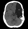
Figure 1
Figure 1
Radiological presentation of an arachnoid cyst (AC). Computed tomography (CT) scan of a large, left-sided temporal AC. Note the splitting of the Sylvian fissure and the compression of the frontal and temporal lobes, all classical features of a Galassi type 3 cyst [10]. The midline is displaced 6 mm to the right. Note the enlargement of the left cranial vault suggesting that the AC was present before the neurocranium was fully developed.
Figure 2
Figure 2
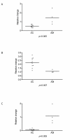
Validation of differentially expressed genes by qRT-PCR. Plots showing the expression of three genes analyzed by quantitative reverse transcriptase real-time polymerase chain reaction (qRT-PCR) for arachnoid cyst tissue (AC, n = 11) and control arachnoid tissue (AM, n = 4) for three genes: A) ATP10D, B) BEND5, and C) SHROOM3. The expression of all three genes was significantly different between the two tissues.
Figure 3
Figure 3
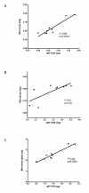
Correlation analysis between microarray signal and quantitative reverse transcriptase real-time polymerase chain reaction (qRT-PCR). Plots showing the correlation between the microarray signal and the expression profile from qRT-PCR analyses for three differentially expressed genes separating arachnoid cysts (AC, n = 7) and arachnoid membranes (AM, n = 2): A) ATP10D, B) BEND5, C) SHROOM3. Data were log transformed before the Pearson correlation was calculated.

Similar articles

Cited by

References

    1. Vernooij MW, Ikram MA, Tanghe HL, Vincent AJ, Hofman A, Krestin GP, Niessen WJ, Breteler MM, Lugt A van der. Incidental findings on brain MRI in the general population. N Engl J Med. 2007;357:1821–1828. doi: 10.1056/NEJMoa070972. - DOI - PubMed
    1. Helland CA, Wester K. A population-based study of intracranial arachnoid cysts: clinical and neuroimaging outcomes following surgical cyst decompression in adults. J Neurol Neurosurg Psychiatry. 2007;78(10):1129–35. - PMC - PubMed
    1. Wester K. Intracranial arachnoid cysts--do they impair mental functions? J Neurol. 2008;255:1113–1120. doi: 10.1007/s00415-008-0011-y. - DOI - PubMed
    1. Wester K. Peculiarities of intracranial arachnoid cysts: location, sidedness, and sex distribution in 126 consecutive patients. Neurosurgery. 1999;45:775–779. doi: 10.1097/00006123-199910000-00008. - DOI - PubMed
    1. Sakai N, Kumagai M, Ueda T, Iwamura M, Nishimura Y, Miwa Y, Shimizu K, Hirata T, Andoh T, Yamada H. [Clinical study on intracranial arachnoid cyst: with reference to the middle cranial fossa] No Shinkei Geka. 1989;17:117–123. - PubMed