. 2010 Feb 26:3:5.
doi: 10.1186/1757-2215-3-5.

Constitutive activation of BMP signalling abrogates experimental metastasis of OVCA429 cells via reduced cell adhesion

Affiliations

Constitutive activation of BMP signalling abrogates experimental metastasis of OVCA429 cells via reduced cell adhesion

Trevor G Shepherd et al. J Ovarian Res. .

Abstract

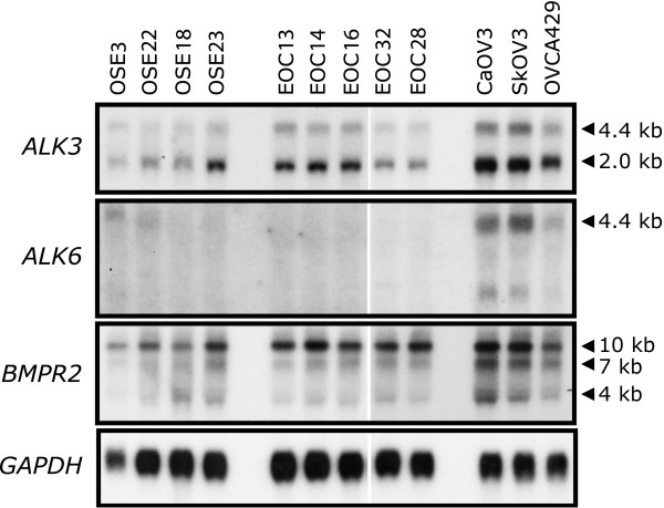
Background: Activation of bone morphogenetic protein (BMP)4 signalling in human ovarian cancer cells induces a number of phenotypic changes in vitro, including altered cell morphology, adhesion, motility and invasion, relative to normal human ovarian surface epithelial cells. From these in vitro analyses, we had hypothesized that active BMP signalling promotes the metastatic potential of ovarian cancer.

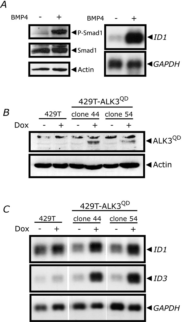
Methods: To test this directly, we engineered OVCA429 human ovarian cancer cells possessing doxycycline-inducible expression of a constitutively-active mutant BMP receptor, ALK3QD, and administered these cells to immunocompromised mice. Further characterization was performed in vitro to address the role of activated BMP signalling on the EOC phenotype, with particular emphasis on epithelial-mesenchymal transition (EMT) and cell adhesion.

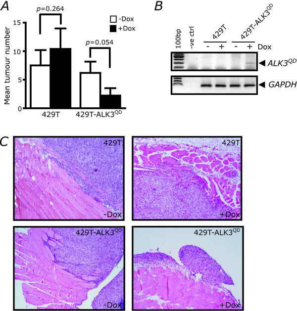
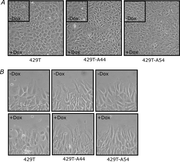
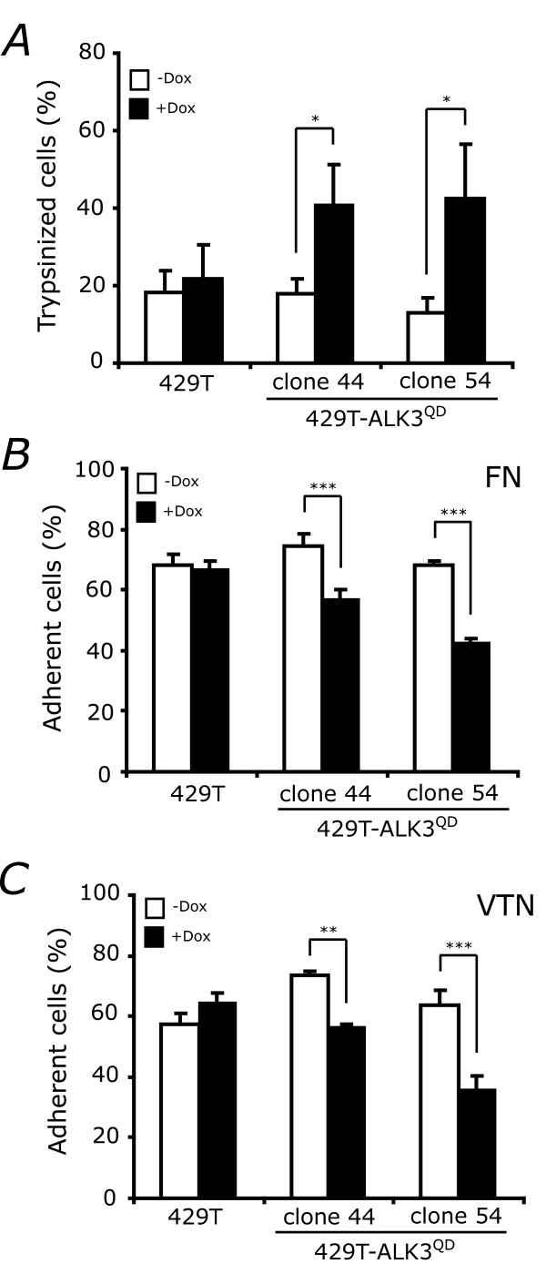
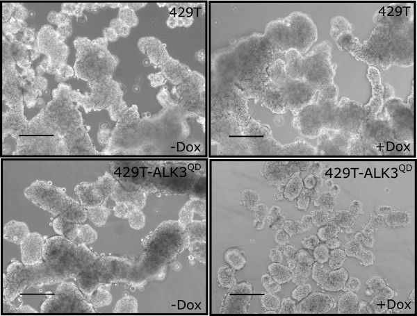
Results: Unexpectedly, doxycycline-induced ALK3QD expression in OVCA429 cells reduced tumour implantation on peritoneal surfaces and ascites formation when xenografted into immunocompromised mice by intraperitoneal injection. To determine the potential mechanisms controlling this in vivo observation, we followed with several cell culture experiments. Doxycycline-induced ALK3QD expression enhanced the refractile, spindle-shaped morphology of cultured OVCA429 cells eliciting an EMT-like response. Using in vitro wound healing assays, we observed that ALK3QD-expressing cells migrated with long, cytoplasmic projections extending into the wound space. The phenotypic alterations of ALK3QD-expressing cells correlated with changes in specific gene expression patterns of EMT, including increased Snail and Slug and reduced E-cadherin mRNA expression. In addition, ALK3QD signalling reduced beta1- and beta3-integrin expression, critical molecules involved in ovarian cancer cell adhesion. The combination of reduced E-cadherin and beta-integrin expression correlates directly with the reduced EOC cell cohesion in spheroids and reduced cell adhesion to the extracellular matrix substrates fibronectin and vitronectin that was observed.

Conclusions: We propose that the key steps of ovarian cancer metastasis, specifically cell cohesion of multicellular aggregates in ascites and cell adhesion for reattachment to secondary sites, may be inhibited by overactive BMP signalling, thereby decreasing the ultimate malignant potential of ovarian cancer in this model system.


Figures

Figure 1
Figure 1
BMP receptor expression in normal human OSE and human ovarian cancer cells. Northern blot analysis was performed on total RNA isolated from early-passage primary cultures of normal human OSE cells (OSE3, OSE18, OSE22, and OSE23), primary human EOC cells (EOC13, EOC14, EOC16, EOC28, and EOC32), and three established human ovarian cancer cell lines (CaOV3, SkOV3, and OVCA429) using probes specific for ALK3, ALK6, and BMPR2 mRNA. ALK3 and BMPR2 mRNA expression was readily detectable (24 h exposure) in all samples analysed, whereas ALK6 mRNA expression was observed only in ovarian cancer cell lines after an extended period of time (8 d exposure). Expression of all three BMP receptors was elevated in the three cell lines versus the primary cultures; however, there was no mean difference in receptor expression between primary cultures of OSE and EOC cells.
Figure 2
Figure 2
Generation of human ovarian cancer OVCA429 cells with Dox-inducible expression of the constitutively-active mutant ALK3 receptor (ALK3QD). (A) Phosphorylation of Smad1 (P-Smad1) and expression of ID1 mRNA are induced in serum-starved OVCA429 cells treated with 10 ng/mL BMP4 for 30 min and 90 min, respectively. Total Smad1 and actin were used as protein loading controls, and GAPDH for RNA loading control. (B) Expression of HA-tagged constitutively-active ALK3 receptor (ALK3QD) was observed by Western analysis in two independent 429T-ALK3QD stable cell clones (A44 and A54) after 24 h treatment with Dox, as compared with control cells (T7Hyg4). Actin was used as a control for protein loading. (C) Activated BMP signalling was confirmed by Northern analysis of ID1 and ID3 mRNA expression from 429T-A44 and 429T-A54 cells, and 429T control cells, treated with Dox for 24 h, or left untreated. GAPDH served as a control for RNA loading.
Figure 3
Figure 3
ALK3QD reduces intraperitoneal tumour formation. Female CD-1 nu/nu athymic nude mice were injected intraperitoneally with a suspension of 5 × 10⁵ cells (either 429T-ALK3QD or 429T control cells), resulting in four groups of fifteen mice (each cell line with or without Dox-containing chow). (A) Fewer Dox-treated mice injected with 429T-ALK3QD cells developed detectable tumour lesions throughout the peritoneal cavity when compared with Dox-treated 429T-injected mice. (B) ALK3QD transgene expression in tumours that formed in nude mice was confirmed by RT-PCR analysis of total RNA with human GAPDH mRNA expression serving as a control for xenograft material present in each sample. (C) Tumour specimens isolated from 429T-injected and 429T-ALK3QD-injected mice fed a Dox-containing or normal chow diet were analyzed histologically by H&E staining. Tumour implants from the peritoneal wall were adherent to the surface of smooth muscle in Dox-treated 429T-ALK3QD-injected mice, whereas localized invasion was evident in specimens from the other groups of mice. 100 × original magnification.
Figure 4
Figure 4
ALK3QD signalling induces EMT marker expression and reduces β1-and β3-integrin expression. Quantitative RT-PCR was performed on total RNA isolated from 429T-ALK3QD cells, and 429T control cells, treated with Dox for 2 d, or left untreated. Human gene-specific primers were used to detect mRNA expression of the EMT markers Snail (SNAI1), Slug (SNAI2), and E-cadherin (CDH1). Quantification of expression of beta- integrins [β1 (ITGB1), and β3 (ITGB3)], and the extracellular matrix components, fibronectin (FN1) and vitronectin (VTN) was also performed. Induction of ID1 and ID3 mRNA expression by ALK3QD served as a positive control. GAPDH mRNA expression was used for normalization, and the fold change in mRNA expression was calculated by the ratio of Dox-treated versus untreated cell samples.
Figure 5
Figure 5
ALK3QD signalling induces an EMT-like morphology of OVCA429 cells. (A) Subconfluent monolayer cultures of 429T-A44 and 429T-A54 cells, and 429T control cells, were treated with Dox for 2 d, or left untreated (inset). At confluence, both 429T-A44 and 429T-A54 cells exhibited a greater proportion of refractile, spindle-shaped cells versus 429T control cells. (B) All cells were grown and treated as described above, but after 2 d a scratch wound was generated. Long cytoplasmic projections extend into the wound space in Dox-treated 429T-A44 and 429T-A54 cells as compared with 429T control cells. Photo images were captured at 100 × original magnification.
Figure 6
Figure 6
ALK3QD signalling decreases OVCA429 cell adhesion. (A) ALK3QD expression increases trypsin-sensitivity of OVCA429 cells. Subconfluent monolayer cultures of 429T-ALK3QD cells (clones A44 and A54), and 429T control cells, were treated with Dox for 2 d (+Dox), or left untreated (-Dox). At confluence, cells were gently trypsinized for 2 min, and the number of suspended cells was scored. The final proportion of released cells was significantly higher in Dox-treated 429T-A44 and -A54 cells versus 429T control cells. (B,C) ALK3QD expression decreases the adhesion of OVCA429 cells to the ECM components fibronectin (FN) and vitronectin (VTN). Cells were cultured in the presence or absence of Dox, radiolabelled with 3H-amino acids, trypsinized, counted, and allowed to recover in serum-containing media. Cells were then seeded at 1 × 10⁵ cells per well that were pre-coated with FN and VTN and number of attached cells were quantified. Dox-treated 429T-A44 and -A54 cells had a significantly-reduced ability to attach to both FN and VTN as compared with untreated cells, and the 429T control cells. (*, p < 0.05; **, p < 0.01; ***, p < 0.001)
Figure 7
Figure 7
ALK3QD signalling reduces OVCA429 spheroid formation. 429T-ALK3QD cells and 429T control cells were grown on Ultra-Low Attachment cluster plates for 2 d while treated with Dox or left untreated. ALK3QD-expressing cells produced smaller multicellular aggregates, or spheroids, than untreated 429T-ALK3QD cells and 429T controls. Scale bar = 200 μm.

References

    1. Naora H, Montell DJ. Ovarian cancer metastasis: integrating insights from disparate model organisms. Nat Rev Cancer. 2005;5:355–66. doi: 10.1038/nrc1611. - DOI - PubMed
    1. Shepherd TG, Nachtigal MW. Identification of a putative autocrine bone morphogenetic protein-signaling pathway in human ovarian surface epithelium and ovarian cancer cells. Endocrinology. 2003;144:3306–14. doi: 10.1210/en.2003-0185. - DOI - PubMed
    1. Nohe A, Keating E, Knaus P, Peterson NO. Signal transduction of bone morphogenetic protein receptors. Cell Signalling. 2004;16:291–9. doi: 10.1016/j.cellsig.2003.08.011. - DOI - PubMed
    1. Hartung A, Bitton-Worms K, Rechtman MM, Wenzel V, Boergermann JH, Hassel S, Henis YI, Knaus P. Different routes of bone morphogenic protein (BMP) receptor endocytosis influence BMP signaling. Mol Cell Biol. 2006;26:7791–805. doi: 10.1128/MCB.00022-06. - DOI - PMC - PubMed
    1. Attisano L, Wrana JL. Smads as transcriptional co-modulators. Curr Opin Cell Biol. 2000;12:235–43. doi: 10.1016/S0955-0674(99)00081-2. - DOI - PubMed