

In vivo lens deficiency of the R49C alphaA-crystallin mutant
- PMID: 20188090
- PMCID: PMC2873126
- DOI: 10.1016/j.exer.2010.02.009
In vivo lens deficiency of the R49C alphaA-crystallin mutant
Abstract
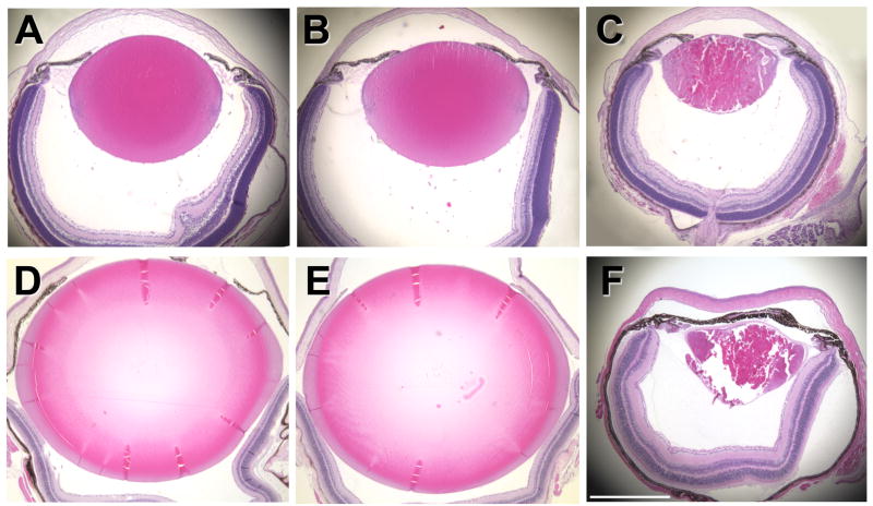
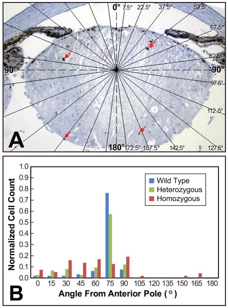
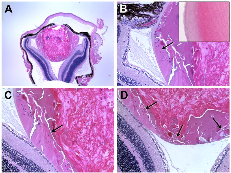
The R49C mutation of alphaA-crystallin (alphaA-R49C) causes hereditary cataracts in humans; patients in a four-generation Caucasian family were found be heterozygous for this autosomal dominant mutation. We previously generated knock-in mouse models of this mutation and found that by 2 months of age, heterozygous mutant mice exhibited minor lens defects including reduced protein solubility, altered signaling in epithelial and fiber cells, and aberrant interactions between alphaA-crystallin and other lens proteins. In contrast, homozygous mutant alphaA-R49C knock-in mice displayed earlier and more extensive lens defects including small eyes and small lenses at birth, death of epithelial and fiber cells, and the formation of posterior, nuclear, and cortical cataracts in the first month of life. We have extended this study to now show that in alphaA-R49C homozygous mutant mice, epithelial cells failed to form normal equatorial bow regions and fiber cells continued to die as the mice aged, resulting in a complete loss of lenses and overall eye structure in mice older than 4 months. These results demonstrate that expression of the hereditary R49C mutant of alphaA-crystallin in vivo is sufficient to adversely affect lens growth, lens cell morphology, and eye function. The death of fiber cells caused by this mutation may ultimately lead to loss of retinal integrity and blindness.
Copyright 2010 Elsevier Ltd. All rights reserved.
Figures
Similar articles
-
Autophagy and UPR in alpha-crystallin mutant knock-in mouse models of hereditary cataracts.Biochim Biophys Acta. 2016 Jan;1860(1 Pt B):234-9. doi: 10.1016/j.bbagen.2015.06.001. Epub 2015 Jun 11. Biochim Biophys Acta. 2016. PMID: 26071686 Free PMC article.
-
AlphaA-crystallin R49Cneo mutation influences the architecture of lens fiber cell membranes and causes posterior and nuclear cataracts in mice.BMC Ophthalmol. 2009 Jul 20;9:4. doi: 10.1186/1471-2415-9-4. BMC Ophthalmol. 2009. PMID: 19619312 Free PMC article.
-
Mechanism of small heat shock protein function in vivo: a knock-in mouse model demonstrates that the R49C mutation in alpha A-crystallin enhances protein insolubility and cell death.J Biol Chem. 2008 Feb 29;283(9):5801-14. doi: 10.1074/jbc.M708704200. Epub 2007 Dec 5. J Biol Chem. 2008. PMID: 18056999
-
Genetics of crystallins: cataract and beyond.Exp Eye Res. 2009 Feb;88(2):173-89. doi: 10.1016/j.exer.2008.10.011. Epub 2008 Nov 1. Exp Eye Res. 2009. PMID: 19007775 Review.
-
Effects of alpha-crystallin on lens cell function and cataract pathology.Curr Mol Med. 2009 Sep;9(7):887-92. doi: 10.2174/156652409789105598. Curr Mol Med. 2009. PMID: 19860667 Review.
Cited by
-
Autophagy and UPR in alpha-crystallin mutant knock-in mouse models of hereditary cataracts.Biochim Biophys Acta. 2016 Jan;1860(1 Pt B):234-9. doi: 10.1016/j.bbagen.2015.06.001. Epub 2015 Jun 11. Biochim Biophys Acta. 2016. PMID: 26071686 Free PMC article.
-
Activation of the unfolded protein response by a cataract-associated αA-crystallin mutation.Biochem Biophys Res Commun. 2010 Oct 15;401(2):192-6. doi: 10.1016/j.bbrc.2010.09.023. Epub 2010 Sep 15. Biochem Biophys Res Commun. 2010. PMID: 20833134 Free PMC article.
-
In vivo substrates of the lens molecular chaperones αA-crystallin and αB-crystallin.PLoS One. 2014 Apr 23;9(4):e95507. doi: 10.1371/journal.pone.0095507. eCollection 2014. PLoS One. 2014. PMID: 24760011 Free PMC article.
-
Changes in relative histone abundance and heterochromatin in αA-crystallin and αB-crystallin knock-in mutant mouse lenses.BMC Res Notes. 2020 Jul 2;13(1):315. doi: 10.1186/s13104-020-05154-7. BMC Res Notes. 2020. PMID: 32616056 Free PMC article.
-
Alpha-crystallin mutations alter lens metabolites in mouse models of human cataracts.PLoS One. 2020 Aug 24;15(8):e0238081. doi: 10.1371/journal.pone.0238081. eCollection 2020. PLoS One. 2020. PMID: 32833997 Free PMC article.
References
Publication types
MeSH terms
Substances
Grants and funding
LinkOut - more resources
Full Text Sources
Medical
Molecular Biology Databases
