. 2010 Feb 23;28(8):1962-74.
doi: 10.1016/j.vaccine.2009.10.099.

Repeated DNA therapeutic vaccination of chronically SIV-infected macaques provides additional virological benefit

Affiliations

Repeated DNA therapeutic vaccination of chronically SIV-infected macaques provides additional virological benefit

Antonio Valentin et al. Vaccine. .

Abstract

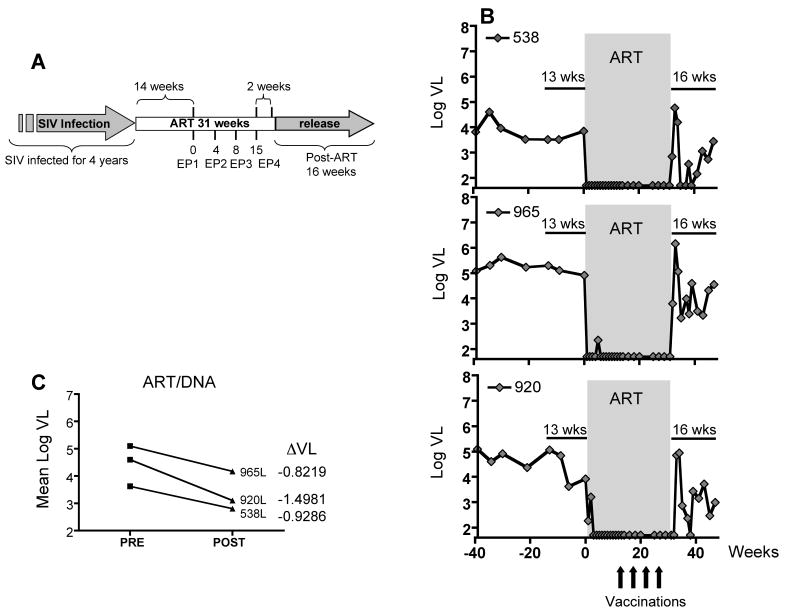
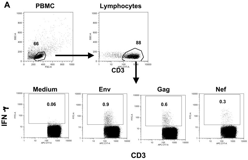
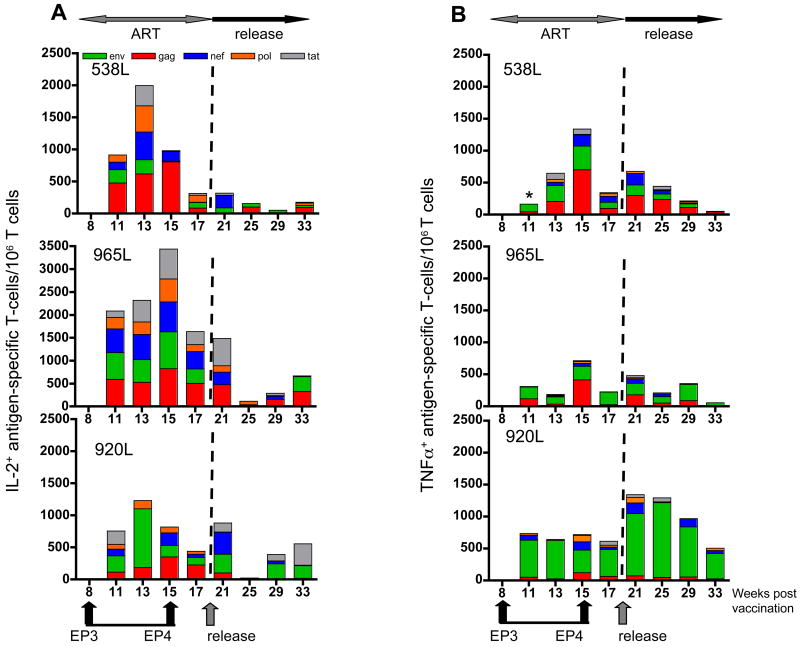
We have previously reported that therapeutic immunization by intramuscular injection of optimized plasmid DNAs encoding SIV antigens effectively induces immune responses able to reduce viremia in antiretroviral therapy (ART)-treated SIVmac251-infected Indian rhesus macaques. We subjected such therapeutically immunized macaques to a second round of therapeutic vaccination using a combination of plasmids expressing SIV genes and the IL-15/IL-15 receptor alpha as molecular adjuvant, which were delivered by the more efficacious in vivo constant-current electroporation. A very strong induction of antigen-specific responses to Gag, Env, Nef, and Pol, during ART (1.2-1.6% of SIV-specific T cells in the circulating T lymphocytes) was obtained with the improved vaccination method. Immunological responses were characterized by the production of IFN-gamma, IL-2, and TNF-alpha either alone, or in combination as double or triple cytokine positive multifunctional T cells. A significant induction of CD4(+) T cell responses, mainly targeting Gag, Nef, and Pol, as well as of CD8(+) T cells, mainly targeting Env, was found in both T cells with central memory and effector memory markers. After release from ART, the animals showed a virological benefit with a further approximately 1 log reduction in viremia. Vaccination with plasmid DNAs has several advantages over other vaccine modalities, including the possibility for repeated administration, and was shown to induce potent, efficacious, and long-lasting recall immune responses. Therefore, these data support the concept of adding DNA vaccination to the HAART regimen to boost the HIV-specific immune responses.


Figures

FIG. 1
FIG. 1
Virological benefit from 2nd round of ART/DNA. (A) Study Outline. SIV-infected macaques were subjected to a 2nd round of ART/DNA after more than 3 years of infection. Previously, we reported the data obtained from the 1st cycle of ART/DNA (von Gegerfelt et al., 2007). After 3.1 to 3.8 years, the animals were subjected to a 2nd round of ART/DNA. The duration of the 2nd ART/DNA was 31 weeks and the animals were vaccinated by in vivo electroporation four times (EP1 to EP4) initiated at week 14 of ART. The animals were released from ART and monitored for another 16 weeks. (B) Viral loads of the three macaques during 2nd round of ART/DNA. Viral load data are shown for 39 weeks prior to ART (PRE), 31 weeks of ART/DNA and 16 weeks post release from ART (POST). (C) Mean virus loads and decrease in mean virus load (ΔVL) comparing 13 weeks PRE and 16 weeks POST treatment. The differences are statistically significant (paired T-test).
FIG. 2
FIG. 2
Increased SIV-specific IFN-γ producing T-cells in DNA immunized macaques during ART. (A) Identification of IFN-γ producing antigen-specific T cells by flow cytometry. PBMC stimulated with different peptide pools of SIV antigens were stained with a cocktail of cell surface antibodies as described in Material and Methods. The main lymphocyte population was gated based on forward and side scatter, and T cells were identified according to CD3 staining. Dot plots show the frequency of IFN-γ+ T cells upon stimulation with Env, Gag or Nef peptide pools. The background of the assay in the presence of medium alone is shown (data is from macaque 965L at week 3 post EP3). (B) The numbers of IFN-γ producing T cells of the three animals after stimulation with Env, Gag, Nef, Pol and Tat peptide pools, respectively, expressed per million circulating T lymphocytes are shown during ART/DNA and after release from ART.
FIG. 2
FIG. 2
Increased SIV-specific IFN-γ producing T-cells in DNA immunized macaques during ART. (A) Identification of IFN-γ producing antigen-specific T cells by flow cytometry. PBMC stimulated with different peptide pools of SIV antigens were stained with a cocktail of cell surface antibodies as described in Material and Methods. The main lymphocyte population was gated based on forward and side scatter, and T cells were identified according to CD3 staining. Dot plots show the frequency of IFN-γ+ T cells upon stimulation with Env, Gag or Nef peptide pools. The background of the assay in the presence of medium alone is shown (data is from macaque 965L at week 3 post EP3). (B) The numbers of IFN-γ producing T cells of the three animals after stimulation with Env, Gag, Nef, Pol and Tat peptide pools, respectively, expressed per million circulating T lymphocytes are shown during ART/DNA and after release from ART.
FIG. 3
FIG. 3
Flow cytometric analysis of T cell subsets. (A) Identification of the subsets of CD4+ and CD8+ T cells. The T cells were divided in central memory (CM) and effector memory (EM) cells based on the pattern of CD28 and CD45RA expression: CD3+CD28+CD45RA- for CM T cells and CD3+CD28- for EM T cells. The two subsets of antigen experienced T cells were further divided in CD4+ and CD8+ populations. Cells with an EM phenotype were mainly CD8+ T cells (91% in macaque 965L), while the majority of CM cells were CD4+ T cells. (B) Identification of SIV-specific IFN-γ+ CM and EM T cells by flow cytometry. Dot plots show the presence of CD4+ and CD8+ T cells with CM and EM markers producing IFN-γ in the presence of Gag and Env SIV peptide pools or medium alone. Numbers inside the gates represent the percentage of IFN-γ+ T cells within the respective parent population. The data shown were obtained from the same animal 965L (3 weeks post EP3) as used in Fig. 3A.
FIG. 3
FIG. 3
Flow cytometric analysis of T cell subsets. (A) Identification of the subsets of CD4+ and CD8+ T cells. The T cells were divided in central memory (CM) and effector memory (EM) cells based on the pattern of CD28 and CD45RA expression: CD3+CD28+CD45RA- for CM T cells and CD3+CD28- for EM T cells. The two subsets of antigen experienced T cells were further divided in CD4+ and CD8+ populations. Cells with an EM phenotype were mainly CD8+ T cells (91% in macaque 965L), while the majority of CM cells were CD4+ T cells. (B) Identification of SIV-specific IFN-γ+ CM and EM T cells by flow cytometry. Dot plots show the presence of CD4+ and CD8+ T cells with CM and EM markers producing IFN-γ in the presence of Gag and Env SIV peptide pools or medium alone. Numbers inside the gates represent the percentage of IFN-γ+ T cells within the respective parent population. The data shown were obtained from the same animal 965L (3 weeks post EP3) as used in Fig. 3A.
FIG. 4
FIG. 4
Comparison of SIV-specific IFN-γ producing T cell subsets induced by immunization during ART. Frequency of SIV-specific CD4+ and CD8+ T cells with central memory (CM) or effector memory (EM) markers were determined as outlined in Fig. 4. Numbers indicate IFN-γ producing T cells after stimulation with Gag (A), Env (B), Nef (C), and Pol (D) peptide pools, expressed per million circulating T lymphocytes.
FIG. 4
FIG. 4
Comparison of SIV-specific IFN-γ producing T cell subsets induced by immunization during ART. Frequency of SIV-specific CD4+ and CD8+ T cells with central memory (CM) or effector memory (EM) markers were determined as outlined in Fig. 4. Numbers indicate IFN-γ producing T cells after stimulation with Gag (A), Env (B), Nef (C), and Pol (D) peptide pools, expressed per million circulating T lymphocytes.
FIG. 5
FIG. 5
Flow cytometric analysis of SIV-specific memory T cell responses using different surface markers. This analysis shows that the frequency and phenotype of the antigen specific cells and of the memory T cell subsets are similar irrespective of the use of the combination of CD28 and CD45RA or CD28 and CD95 markers to define these T cell populations. Briefly, PBMC of macaque M538 (at 7 weeks post EP3) were analyzed by flow cytometry after peptide (env) stimulation and staining with monoclonal antibodies against different sets of markers as described in Materials and Methods. (A) The main lymphocyte population was gated based on forward and side scatter, and T cells were identified according to CD3 staining. These T cells were classified as effector memory (EM) and central memory (CM) cells based in the pattern of staining with CD28 and CD45RA (EM1 and CM1) or CD28 and CD95 (EM2 and CM2) (lower plots). (B) Phenotypic analysis of the antigen-specific (IFN-γ+) T cells. The frequency of Env-specific T cells was determined based on IFN-γ production. The cells were classified as EM1 and EM2 and CM1 and CM2 T cells according to the expression of either CD28 and CD45RA or CD28 and CD95, respectively, and the frequency of CD4+ and CD8+ T cells within each of these populations was determined. (C) Analysis of the frequency of IFN-γ+ Env-specific T cells, as well as the CD4+ and CD8+ distribution within the memory subsets as defined in panel A.
FIG. 6
FIG. 6
Induction of SIV-specific IL-2- and TNFα-producing T cells induced by DNA vaccination during ART. The analysis was performed using similar strategy as described for Fig. 3. IL-2 (A) and TNFα (B) producing T cells after stimulation with Env, Gag, Nef, Pol, and Tat peptide pools, respectively, were expressed per million circulating T lymphocytes. The ART and release periods are indicated. *, Tat-specific immune response was not determined.
FIG. 7
FIG. 7
Induction of dual cytokine positive SIV-specific T cells in vaccinated animals. The dual IFN-γ plus IL-2 (A) and IFN-γ plus TNFα (B) producing SIV-specific (Gag, Env, Nef) T cells are indicated.
FIG. 8
FIG. 8
Induction of multifunctional SIV-specific T cells. A comparison of the levels of double positive (from Fig. 7; light grey IFNγ+IL-2+; dark grey and IFNγ+ TNFα+) and triple positive (IFNγ+ IL-2+ TNFα+; striped bar) total SIV specific T cells is shown.
FIG. 9
FIG. 9
Humoral immune responses during ART and DNA vaccination and the period after release from ART. The presence of binding antibodies to Env (gp120) (A) and to Gag p27 (B) was measured in plasma prior, during and after therapy.

Similar articles

Cited by

References

    1. Aberle H, Bauer A, Stappert J, Kispert A, Kemler R. beta-catenin is a target for the ubiquitin-proteasome pathway. EMBO J. 1997;16:3797–3804. - PMC - PubMed
    1. Aihara H, Miyazaki J. Gene transfer into muscle by electroporation in vivo. Nat Biotechnol. 1998;16:867–870. - PubMed
    1. Andrieu JM, Lu W. A dendritic cell-based vaccine for treating HIV infection: background and preliminary results. J Intern Med. 2007;261:123–131. - PubMed
    1. Bansal A, Jackson B, West K, Wang S, Lu S, Kennedy JS, Goepfert PA. Multifunctional T-cell characteristics induced by a polyvalent DNA prime/protein boost human immunodeficiency virus type 1 vaccine regimen given to healthy adults are dependent on the route and dose of administration. J Virol. 2008;82:6458–6469. - PMC - PubMed
    1. Bergamaschi C, Rosati M, Jalah R, Valentin A, Kulkarni V, Alicea C, Zhang GM, Patel V, Felber BK, Pavlakis GN. Intracellular interaction of interleukin IL-15 with its receptor alpha during production leads to mutual stabilization and increased bioactivity. J Biol Chem. 2007;283:4189–4199. - PubMed

Publication types

MeSH terms