Case Reports
. 2010 Mar 12;86(3):389-98.
doi: 10.1016/j.ajhg.2010.01.034. Epub 2010 Feb 25.

Homozygosity for a missense mutation in SERPINH1, which encodes the collagen chaperone protein HSP47, results in severe recessive osteogenesis imperfecta

Affiliations
Case Reports

Homozygosity for a missense mutation in SERPINH1, which encodes the collagen chaperone protein HSP47, results in severe recessive osteogenesis imperfecta

Helena E Christiansen et al. Am J Hum Genet. .

Abstract

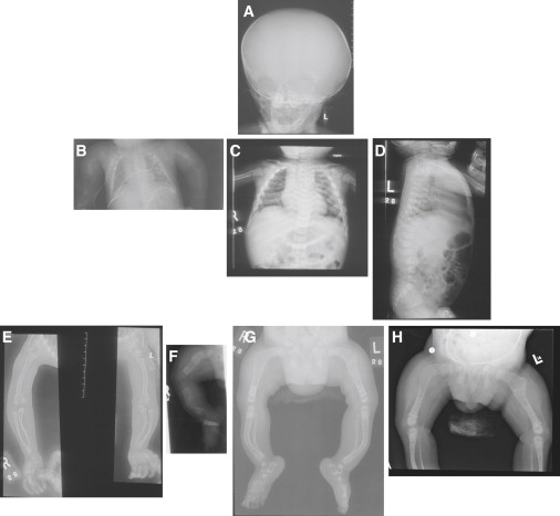
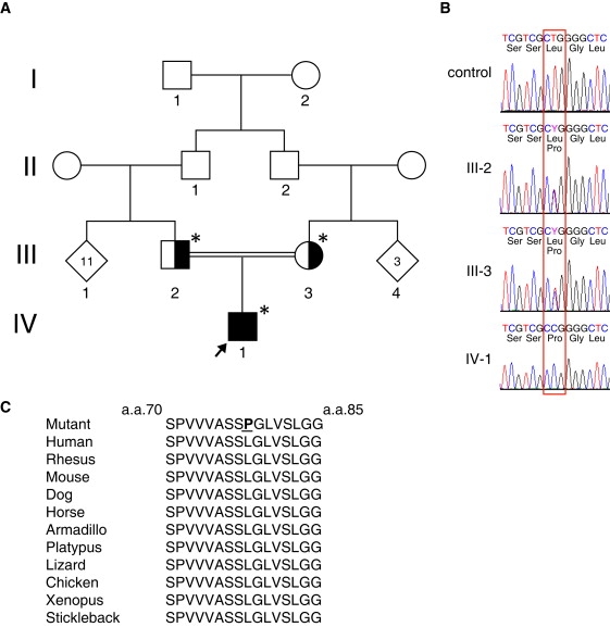
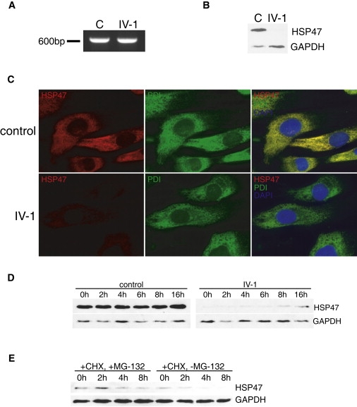
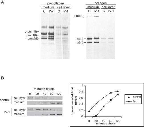
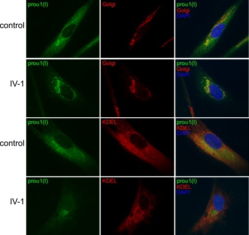
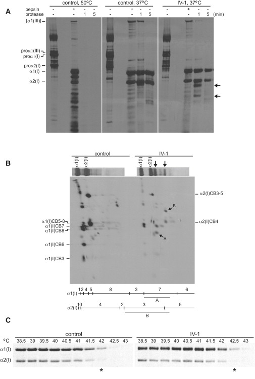
Osteogenesis imperfecta (OI) is characterized by bone fragility and fractures that may be accompanied by bone deformity, dentinogenesis imperfecta, short stature, and shortened life span. About 90% of individuals with OI have dominant mutations in the type I collagen genes COL1A1 and COL1A2. Recessive forms of OI resulting from mutations in collagen-modifying enzymes and chaperones CRTAP, LEPRE1, PPIB, and FKBP10 have recently been identified. We have identified an autosomal-recessive missense mutation (c.233T>C, p.Leu78Pro) in SERPINH1, which encodes the collagen chaperone-like protein HSP47, that leads to a severe OI phenotype. The mutation results in degradation of the endoplasmic reticulum resident HSP47 via the proteasome. Type I procollagen accumulates in the Golgi of fibroblasts from the affected individual and a population of the secreted type I procollagen is protease sensitive. These findings suggest that HSP47 monitors the integrity of the triple helix of type I procollagen at the ER/cis-Golgi boundary and, when absent, the rate of transit from the ER to the Golgi is increased and helical structure is compromised. The normal 3-hydroxylation of the prolyl residue at position 986 of the triple helical domain of proalpha1(I) chains places the role of HSP47 downstream from the CRTAP/P3H1/CyPB complex that is involved in prolyl 3-hydroxylation. Identification of this mutation in SERPINH1 gives further insight into critical steps of the collagen biosynthetic pathway and the molecular pathogenesis of OI.


Figures

Figure 1
Figure 1
X-Rays Radiographs of the skull at age 1 year (A) demonstrate diminished calvarial mineralization and enlargement of the calvarial vault. At birth, the chest (B) was small, the ribs were thin and some had healing fractures, one clavicle was bent, both humeral bones had fractures, platyspondyly was present, and overall mineralization appeared diminished. By 1 year of age (C and D), the ribs had become broad, platyspondyly was still apparent with diminished central mineralization of the vertebral bodies, and the humeral bones had a tubular shape, thin cortices, and diminished mineralization. The deformity and decreased mineralization of the bones in the arms are apparent at 1 year (E), and the minimal effect of pamidronate can be seen. At 1 month (F), 1 year (G), and 28 months (H), there is rhizomelic shortening and limited modeling of the long bones with very thin cortices. The lines of increased mineralization reflect the successive pamidronate infusions.
Figure 2
Figure 2
Identification of a Homozygous Mutation in SERPINH1 (A) Family pedigree with consanguineous heterozygous parents (III-2 and III-3) and affected proband (IV-1, black arrow). Asterisks indicate individuals studied. (B) Homozygous c.233T>C, p.Leu78Pro mutation in exon 1 of SERPINH1 in proband, IV-1 (red box). Parents (III-2 and III-3) were heterozygous at this site. (C) The completely conserved 16 amino acid sequence in HSP47 that contains the leucine-to-proline substitution.
Figure 3
Figure 3
Mutation of SERPINH1 Generates an Unstable Protein (A) PCR amplification of cDNA (35 cycles for each) from control (C) and from patient (IV-1) showed that the mRNA for SERPINH1 was stable. (B) Western blot analysis indicated that HSP47 was significantly decreased in patient cells (IV-1). (C) Immunocytochemistry with antibodies to HSP47 and the ER marker protein disulfide isomerase (PDI) showed a normal ER distribution of HSP47 in control cells and a severe reduction in HSP47 in patient cells (IV-1). The merged image (right column) confirms the absence of staining for HSP47. (D) Western blot showed partial rescue of HSP47 beginning at ∼8 hr of treatment with proteasome inhibitor MG-132. (E) HSP47 was stabilized after treatment of cells with cycloheximide (CHX) and MG-132 but in the absence of MG-132, even with continuous presence of CHX, previously stabilized HSP47 was destroyed.
Figure 4
Figure 4
Type I Procollagen Synthesis and Secretion (A) Labeling of cells with [3H]-proline for 16 hr showed normal synthesis and secretion of type I procollagen into the culture medium and normal electrophoretic mobilities of the constituent chains. The chains of the procollagens were separated under reducing conditions. The collagens represent the procollagens after digestion with pepsin and electrophoresis under nonreducing conditions. (B) Cells were labeled for 1 hr with [3H]-proline then chased for up to 2 hr with unlabeled proline. The secretion of the trimers of type I procollagen showed a delay in the rate of secretion in the patient cells, quantitated in the graph to the right.
Figure 5
Figure 5
Type I Collagen Accumulates in the Golgi of HSP47 Mutant Cells Immunocytochemistry with antibodies against the N-propeptide of proα1(I), Golgi marker 58K Golgi protein, and ER marker KDEL showed localization of type I procollagen to both Golgi and ER in control cells. In patient cells (IV-1 rows), type I procollagen was diminished in the ER and accentuated in the Golgi.
Figure 6
Figure 6
Mutation of HSP47 Affects Protease Sensitivity and Thermostability of the Type I Collagen Triple Helix (A) Proteins in medium from cultured dermal fibroblasts labeled overnight with [3H]-proline were digested for 1 or 5 min with a combination of trypsin and chymotrypsin at 37°C without prior cooling of the sample. Pepsin-treated samples were ethanol precipitated at 4°C and then digested for 2 hr at 16°C. Procollagens secreted into the culture medium by control cells and then pretreated at 50°C for 15 min showed complete degradation after treatment with trypsin/chymotrypsin while procollagens from medium left at 37°C had protease-resistant triple helical domains. A subset of type I procollagen molecules secreted from patient cells were cleaved asymmetrically at 37°C (the black arrows indicate fragments seen in affected cells and either not seen or in low abundance in the control cells). (B) Characterization by cyanogen bromide peptide mapping of the cleavage products of type I procollagen after trypsin/chymotrypsin treatment. The gel lanes from runs equivalent to those in (A) were excised and the bands cleaved with cyanogen bromide. The larger of the new bands, which migrated just below α2(I), was derived from α1(I). The CB7 fragment is missing and replaced by fragment A. The size of the fragment indicates that the parent peptide had been cleaved in CB7 near the mammalian collagenase cleavage site (775–776 in the triple helical domain). The smaller fragment in the parent gel was derived from α2(I). The CB3-5 fragment was shortened, fragment B, and the estimated size indicated that it had been cleaved in the region of the collagenase cleavage site. (C) Thermal stability of type I collagen molecules. Cells were incubated overnight with [3H]-proline in the presence of sodium ascorbate (50 μg/ml). The secreted proteins were collected by precipitation with ethanol and then reheated to the temperatures shown at which point they were treated with trypsin/chymotrypsin for 2 min. The thermostability of type I collagen was increased in patient cells by about 0.5°C ().

Similar articles

Cited by

References

    1. Byers P.H., Cole W.G. Osteogenesis imperfecta. In: Royce P.M., Steinmann B., editors. Connective Tissue and It's Heritable Disorders: Molecular, Genetic, and Medical Aspects. Wiley-Liss, Inc.; New York: 2002. pp. 385–430.
    1. Rauch F., Glorieux F.H. Osteogenesis imperfecta. Lancet. 2004;363:1377–1385. - PubMed
    1. Marini J.C., Forlino A., Cabral W.A., Barnes A.M., San Antonio J.D., Milgrom S., Hyland J.C., Körkkö J., Prockop D.J., De Paepe A. Consortium for osteogenesis imperfecta mutations in the helical domain of type I collagen: Regions rich in lethal mutations align with collagen binding sites for integrins and proteoglycans. Hum. Mutat. 2007;28:209–221. - PMC - PubMed
    1. Bodian D.L., Chan T.F., Poon A., Schwarze U., Yang K., Byers P.H., Kwok P.Y., Klein T.E. Mutation and polymorphism spectrum in osteogenesis imperfecta type II: Implications for genotype-phenotype relationships. Hum. Mol. Genet. 2009;18:463–471. - PMC - PubMed
    1. Morello R., Bertin T.K., Chen Y., Hicks J., Tonachini L., Monticone M., Castagnola P., Rauch F., Glorieux F.H., Vranka J. CRTAP is required for prolyl 3-hydroxylation and mutations cause recessive osteogenesis imperfecta. Cell. 2006;127:291–304. - PubMed

Publication types

MeSH terms