

Tailoring the degradation kinetics of poly(ester carbonate urethane)urea thermoplastic elastomers for tissue engineering scaffolds
- PMID: 20188411
- PMCID: PMC2855340
- DOI: 10.1016/j.biomaterials.2010.02.005
Tailoring the degradation kinetics of poly(ester carbonate urethane)urea thermoplastic elastomers for tissue engineering scaffolds
Abstract
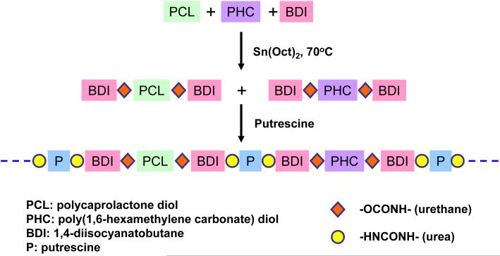
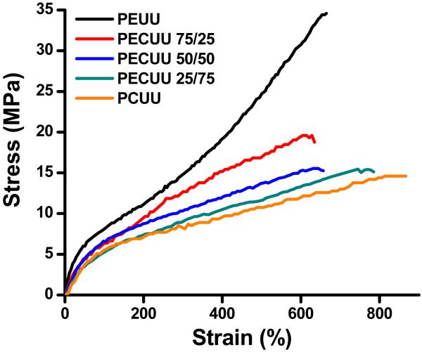
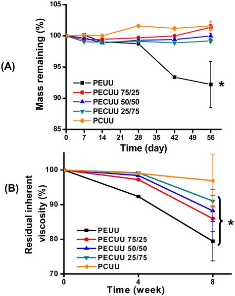
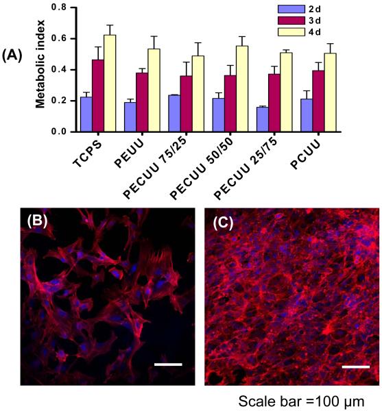
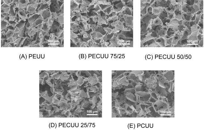
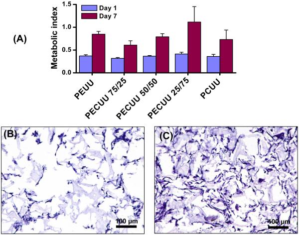
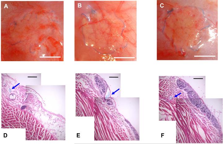
Biodegradable elastomeric scaffolds are of increasing interest for applications in soft tissue repair and regeneration, particularly in mechanically active settings. The rate at which such a scaffold should degrade for optimal outcomes, however, is not generally known and the ability to select from similar scaffolds that vary in degradation behavior to allow such optimization is limited. Our objective was to synthesize a family of biodegradable polyurethane elastomers where partial substitution of polyester segments with polycarbonate segments in the polymer backbone would lead to slower degradation behavior. Specifically, we synthesized poly(ester carbonate)urethane ureas (PECUUs) using a blended soft segment of poly(caprolactone) (PCL) and poly(1,6-hexamethylene carbonate) (PHC), a 1,4-diisocyanatobutane hard segment and chain extension with putrescine. Soft segment PCL/PHC molar ratios of 100/0, 75/25, 50/50, 25/75, and 0/100 were investigated. Polymer tensile strengths varied from 14 to 34 MPa with breaking strains of 660-875%, initial moduli of 8-24 MPa and 100% recovery after 10% strain. Increased PHC content was associated with softer, more distensible films. Scaffolds produced by salt leaching supported smooth muscle cell adhesion and growth in vitro. PECUU in aqueous buffer in vitro and subcutaneous implants in rats of PECUU scaffolds showed degradation slower than comparable poly(ester urethane)urea and faster than poly(carbonate urethane)urea. These slower degrading thermoplastic polyurethanes provide opportunities to investigate the role of relative degradation rates for mechanically supportive scaffolds in a variety of soft tissue repair and reconstructive procedures.
Copyright (c) 2010 Elsevier Ltd. All rights reserved.
Figures
Similar articles
-
Biodegradable polyurethane ureas with variable polyester or polycarbonate soft segments: effects of crystallinity, molecular weight, and composition on mechanical properties.Biomacromolecules. 2011 Sep 12;12(9):3265-74. doi: 10.1021/bm2007218. Epub 2011 Jul 26. Biomacromolecules. 2011. PMID: 21755999 Free PMC article.
-
Synthesis, characterization and surface modification of low moduli poly(ether carbonate urethane)ureas for soft tissue engineering.Acta Biomater. 2009 Oct;5(8):2901-12. doi: 10.1016/j.actbio.2009.04.016. Epub 2009 May 4. Acta Biomater. 2009. PMID: 19433136
-
Preparation and characterization of highly porous, biodegradable polyurethane scaffolds for soft tissue applications.Biomaterials. 2005 Jun;26(18):3961-71. doi: 10.1016/j.biomaterials.2004.10.018. Biomaterials. 2005. PMID: 15626443 Free PMC article.
-
Controllable degradation kinetics of POSS nanoparticle-integrated poly(ε-caprolactone urea)urethane elastomers for tissue engineering applications.Sci Rep. 2015 Oct 14;5:15040. doi: 10.1038/srep15040. Sci Rep. 2015. PMID: 26463421 Free PMC article.
-
Elastomers in vascular tissue engineering.Curr Opin Biotechnol. 2016 Aug;40:149-154. doi: 10.1016/j.copbio.2016.04.008. Epub 2016 May 2. Curr Opin Biotechnol. 2016. PMID: 27149017 Free PMC article. Review.
Cited by
-
Decellularized liver bioscaffold: a histological and immunohistochemical comparison between normal, fibrotic and hepatocellular carcinoma.Clin Exp Hepatol. 2019 Mar;5(1):35-47. doi: 10.5114/ceh.2019.83155. Epub 2019 Feb 20. Clin Exp Hepatol. 2019. PMID: 30915405 Free PMC article.
-
Concomitant control of mechanical properties and degradation in resorbable elastomer-like materials using stereochemistry and stoichiometry for soft tissue engineering.Nat Commun. 2021 Jan 19;12(1):446. doi: 10.1038/s41467-020-20610-5. Nat Commun. 2021. PMID: 33469013 Free PMC article.
-
Nanometer-sized extracellular matrix coating on polymer-based scaffold for tissue engineering applications.J Biomed Mater Res A. 2016 Jan;104(1):94-103. doi: 10.1002/jbm.a.35544. Epub 2015 Aug 6. J Biomed Mater Res A. 2016. PMID: 26194176 Free PMC article.
-
Nitro-Oleic Acid (NO2-OA) Release Enhances Regional Angiogenesis in a Rat Abdominal Wall Defect Model.Tissue Eng Part A. 2018 Jun;24(11-12):889-904. doi: 10.1089/ten.TEA.2017.0349. Epub 2018 Feb 27. Tissue Eng Part A. 2018. PMID: 29187125 Free PMC article.
-
Development and Future Trends of Protective Strategies for Magnesium Alloy Vascular Stents.Materials (Basel). 2023 Dec 22;17(1):68. doi: 10.3390/ma17010068. Materials (Basel). 2023. PMID: 38203922 Free PMC article. Review.
References
-
- Lee SJ, Liu J, Oh SH, Soker S, Atala A, Yoo JJ. Development of a composite vascular scaffolding system that withstands physiological vascular conditions. Biomaterials. 2008;29:2891–8. - PubMed
-
- Webb AR, Yang J, Ameer GA. Biodegradable polyester elastomers in tissue engineering. Expert Opin Biol Ther. 2004;4:801–12. - PubMed
-
- Guelcher SA. Biodegradable polyurethanes: synthesis and applications in regenerative medicine. Tissue Eng Part B Rev. 2008;14:3–17. - PubMed
-
- Wang YD, Ameer GA, Sheppard BJ, Langer R. A tough biodegradable elastomer. Nat Biotechnol. 2002;20:602–6. - PubMed
Publication types
MeSH terms
Substances
Grants and funding
LinkOut - more resources
Full Text Sources
Other Literature Sources
