. 2010 May 4;9(5):478-87.
doi: 10.1016/j.dnarep.2010.01.011. Epub 2010 Feb 25.

Interaction between human mismatch repair recognition proteins and checkpoint sensor Rad9-Rad1-Hus1

Affiliations

Interaction between human mismatch repair recognition proteins and checkpoint sensor Rad9-Rad1-Hus1

Haibo Bai et al. DNA Repair (Amst). .

Abstract

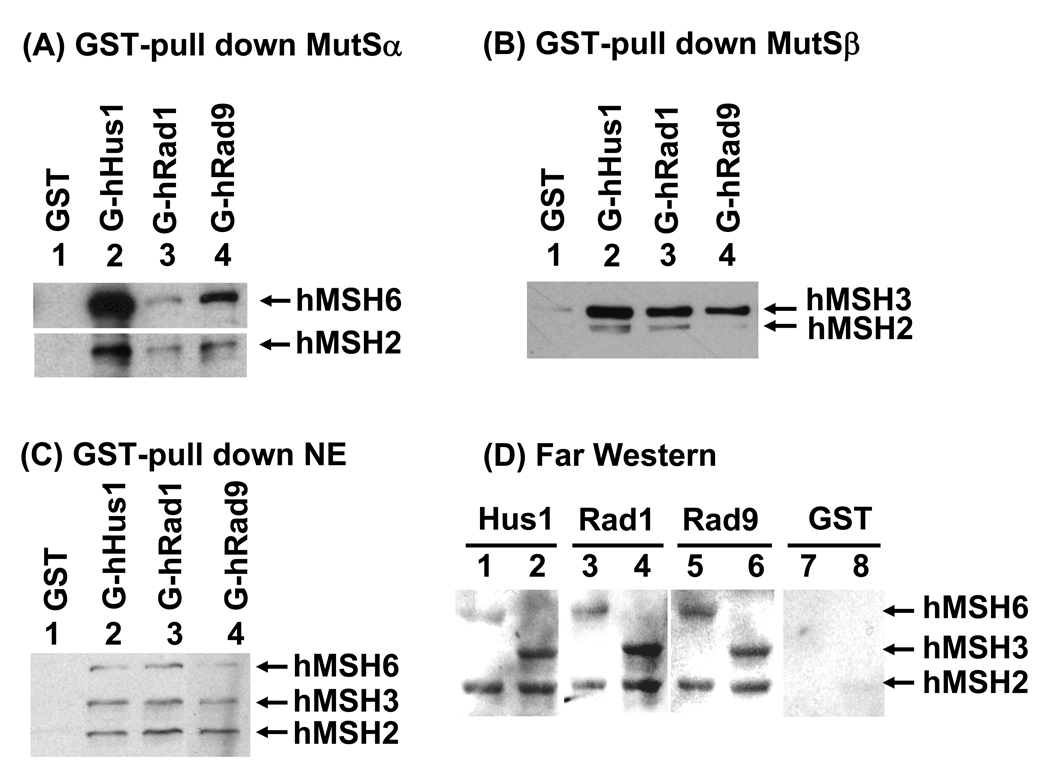
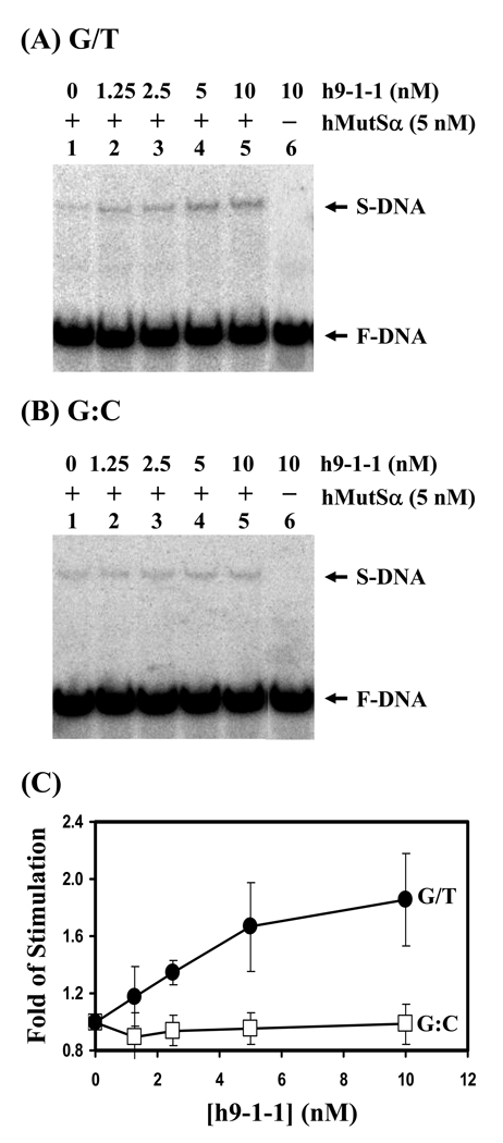
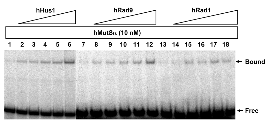
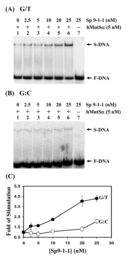
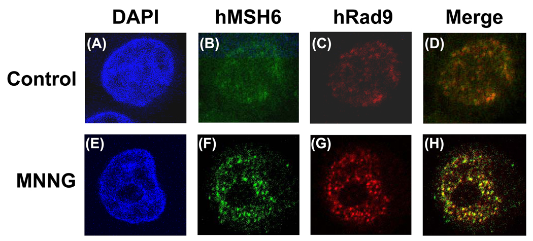
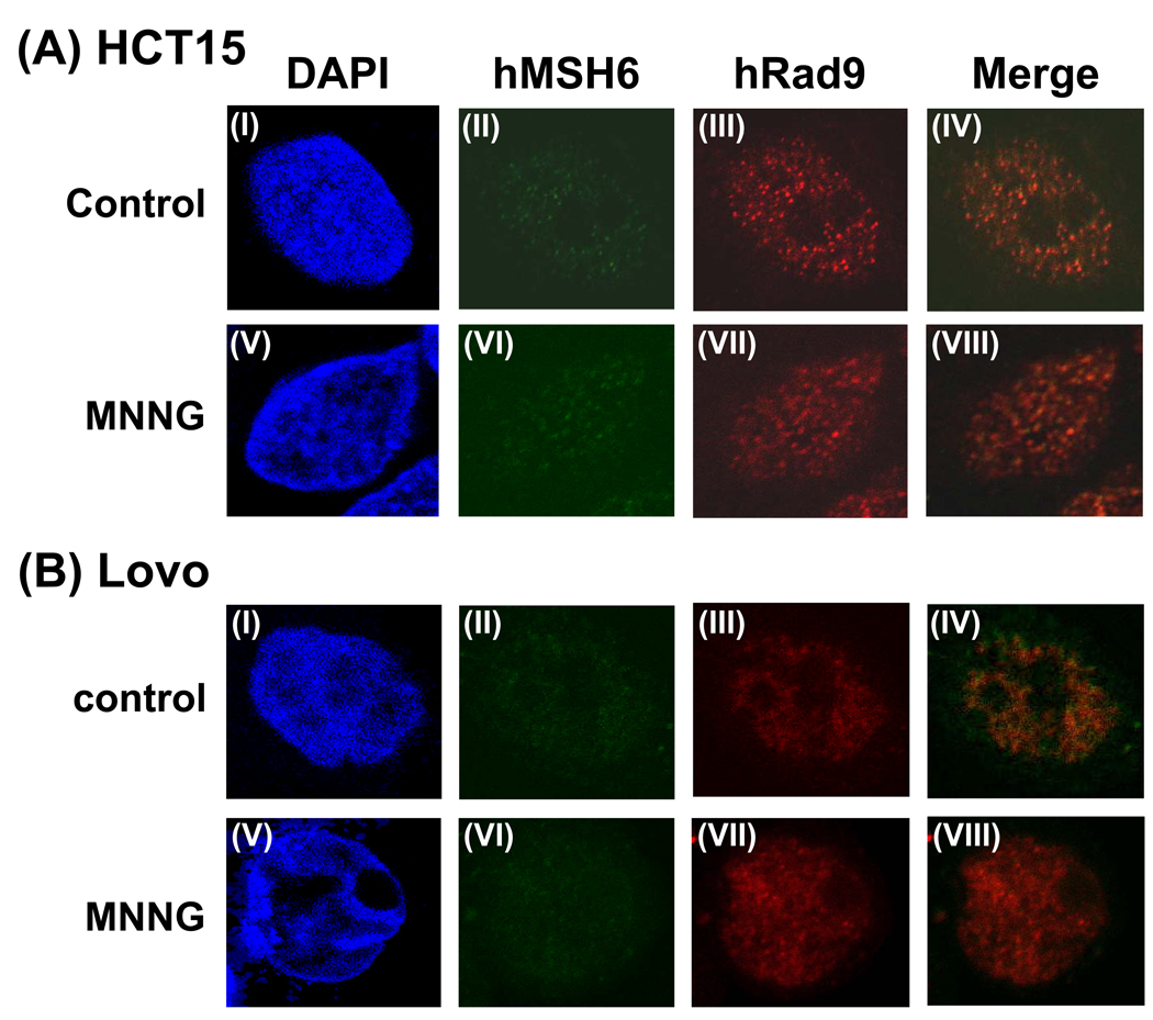
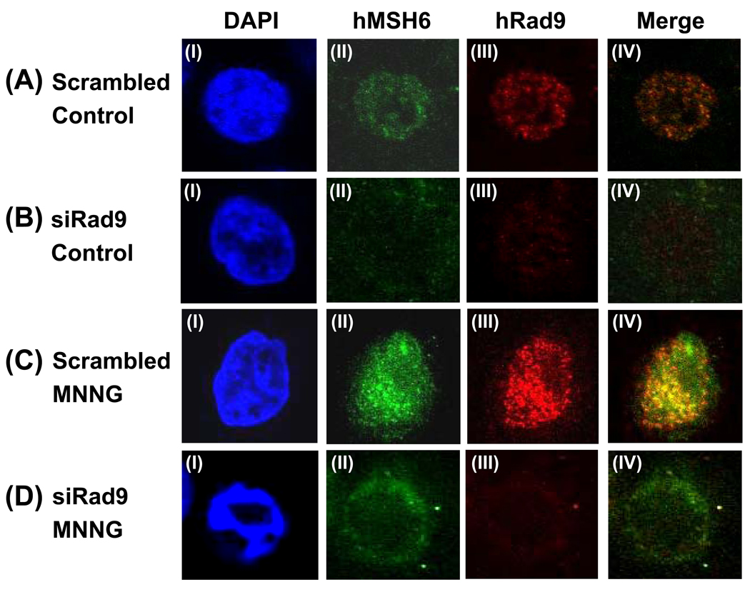
In eukaryotic cells, the cell cycle checkpoint proteins Rad9, Rad1, and Hus1 form the 9-1-1 complex which is structurally similar to the proliferating cell nuclear antigen (PCNA) sliding clamp. hMSH2/hMSH6 (hMutS alpha) and hMSH2/hMSH3 (hMutS beta) are the mismatch recognition factors of the mismatch repair pathway. hMutS alpha has been shown to physically and functionally interact with PCNA. Moreover, DNA methylating agent N-methyl-N'-nitro-N-nitrosoguanidine (MNNG) treatment induces the G2/M cell cycle arrest that is dependent on the presence of hMutS alpha and hMutL alpha. In this study, we show that each subunit of the human 9-1-1 complex physically interacts with hMSH2, hMSH3, and hMSH6. The 9-1-1 complex from both humans and Schizosaccharomyces pombe can stimulate hMutS alpha binding with G/T-containing DNA. Rad9, Rad1, and Hus1 individual subunits can also stimulate the DNA binding activity of hMutS alpha. Human Rad9 and hMSH6 colocalize to nuclear foci of HeLa cells after exposure to MNNG. However, Rad9 does not form foci in MSH6 defective cells following MNNG treatment. In Rad9 knockdown untreated cells, the majority of the MSH6 is in cytoplasm. Following MNNG treatment, Rad9 knockdown cells has abnormal nuclear morphology and MSH6 is distributed around nuclear envelop. Our findings suggest that the 9-1-1 complex is a component of the mismatch repair involved in MNNG-induced damage response.


Figures

Fig. 1
Fig. 1
Physical interactions between human 9-1-1 subunits and hMutSα and hMutSβ. (A) Pull-down of purified hMutSα by immobilized GST-hHus1, GST-hRad1 and GST-hRad9. hMutSα (450 ng) purified from baculovirus expression system was incubated with 300 ng of GST-hHus1, GST-hRad1, GST-hRad9 or GST alone immobilized on glutathione Sepharose 4B. The pellets were fractionated by 8% SDS-polyacrylamide gel followed by Western blot analysis with antibodies against hMSH2 and hMSH6. The exposure time for the blot with hMSH6 antibody was different from the one with hMSH2 antibody. (B) Pull-down of purified hMutSβ by immobilized GST-hHus1, GST-hRad1 and GST-hRad9. The procedures are similar as described in (A) except that hMutSβ (400 ng) was used and Western blot analysis was performed with antibodies against hMSH2 and hMSH3. (C) Pull-down of hMutSα and hMutSβ from HeLa cell extracts by immobilized GST-hHus1, GST-hRad1 and GST-hRad9. Similar experiments were performed as those in (A) but with 750 µg HeLa cell extracts instead of purified hMutSα and Western blot analysis was performed with antibodies against hMSH2, hMSH3 and hMSH6. (D) Far-Western analysis. Purified hMutSα (hMSH2/hMSH6) (odd lanes) and hMutSβ (hMSH2/hMSH3) (even lanes) (10 pmol each) were separated by 8% SDS-polyacrylamide gel and transferred onto a nitrocellulose membrane. The membrane was incubated with E. coli extracts containing hHus1, hRad1, hRad9, or GST as indicated. Subsequent Western blot analysis was performed with antibodies against GST.
Fig. 2
Fig. 2
Human 9-1-1 complex stimulates hMutSα DNA binding activity. (A) hMutSα binding with G/T-containing DNA was stimulated by h9-1-1 complex. Lane 1, DNA substrates were incubated with 5 nM purified hMutSα. Lanes 2-5, reactions are similar to lane 1 but with increasing amounts of h9-1-1 complex (1.25, 2.5, 5, and 10 nM, respectively). Lane 6, DNA substrates were incubated with 10 nM h9-1-1 complex only. The arrows mark the free DNA substrate (F-DNA) and hMutSα-DNA complex (S-DNA). (B) hMutSα binding with homoduplex was not affected by h9-1-1 complex. The binding reactions were similar with those in (A) but with homoduplex (G:C) substrates. (C) Quantitative analyses of fold stimulation of h9-1-1 complex on hMutSα binding activity with G/T-containing DNA (closed circles) and homoduplex (opened squares) from three experiments. The fold of stimulation was calculated by dividing the percentage of DNA-protein complex in the presence of the 9-1-1 complex by the percentage of bound DNA in the absence of the 9-1-1 complex. The error bars reported are the standard deviations of the averages.
Fig. 3
Fig. 3
hMutSα binding activity with G/T-DNA can be stimulated by hHus1, hRad9, and hRad1. Reactions are similar to Fig. 2 but with 10 nM purified hMutSα and increasing amounts of hHus1, Rad9, and Rad1 (15.6, 31.2, 62.5, 125, and 250 nM, respectively). At 6-fold molar excess over hMutSα, hHus1, hRad1, and hRad9 could stimulate the binding ability of hMutSα to G/T mismatches by 3.8, 2.7, and 1.8-fold, respectively.
Fig. 4
Fig. 4
S. pombe 9-1-1 complex can stimulate hMutSα binding activity. (A) hMutSα binding activity with G/T-containing DNA was stimulated by Sp9-1-1 complex. Reactions are similar to Fig. 2. Lane 1, DNA substrates were incubated with 5 nM purified hMutSα. Lanes 2-6, reactions are similar to lane 1 but with increasing amounts of Sp9-1-1 complex (2.5, 5, 10, 20, and 25 nM, respectively). Lane 7, DNA substrates were incubated with 25 nM Sp9-1-1 complex only. (B) hMutSα binding activity with homoduplex was stimulated by Sp9-1-1 complex to a less extent. The experiments were similar with those in (A) but with homoduplex (G:C) substrates. (C) Quantitative analyses of fold stimulation of Sp9-1-1 complex on hMutSα binding activity with G/T-containing DNA (closed circles) and homoduplex (opened squares) from three experiments. The fold of stimulation was calculated by dividing the percentage of DNA-protein complex in the presence of the 9-1-1 complex by the percentage of bound DNA in the absence of the 9-1-1 complex. The error bars reported are the standard deviations of the averages.
Fig. 5
Fig. 5
Colocalization of hMSH6 and hRad9 in MNNG-induced nuclear foci as determined by immunofluorescence. HeLaS3 cells were untreated (upper panel) or treated with 10 µM MNNG in the presence of O6-BG (see Material and Methods) (lower panel). The cells were immunostained with antibody against hMSH6 (green, B and F) and anti-hRad9 antibody (Red, C and G). A and E, DAPI-stained nuclei. D is the merge image of B and C. H is the merge image of F and G. Colocalization of hMSH6 and hRad9 foci is visualized as yellow spots.
Fig. 6
Fig. 6
No hRad9 foci formation in MMR defective HCT15 and LoVo cells following MNNG treatment. (A) HCT15 cells which contain MSH6 frame-shift mutation were untreated (upper panel) or treated with 10 µM MNNG in the presence of O6-BG (lower panel). Immunofluorence staining was performed similarly to Fig. 5. (B) LoVo cells which contain a large deletion in MSH2 gene and unstable MSH6 were untreated (upper panel) or treated with 10 µM MNNG in the presence of O6-BG (lower panel). Immunofluorence staining was performed similarly to Fig. 5. IV is the merge image of II and III. VIII is the merge image of VI and VII.
Fig. 7
Fig. 7
Altered hMSH6 distribution in hRad9 knockdown cells. (A) Untreated HeLa cells transfected with scrambled siRNA. (B) Untreated HeLa cells transfected with hRad9 specific siRNA. (C) HeLa cells transfected with scrambled siRNA were treated with 10 µM MNNG in the presence of O6-BG. (D) HeLa cells transfected with hRad9 specific siRNA were treated with 10 µM MNNG in the presence of O6-BG. Immunofluorence staining was performed similarly to Fig. 5. IV is the merge image of II and III. The expression of hRad9 is much reduced in (B, III) and (D, III). The majority of the hMSH6 was observed in cytoplasm (B, II) in untreated cells and was distributed around the outside of the nuclear envelop in MNNG treated cells (D, II). DAPI staining indicated that Rad9 knockdown cells has abnormal nuclear morphology following MNNG treatment (D, I).

References

    1. Bartek J, Lukas C, Lukas J. Checking on DNA damage in S phase. Nat. Rev. Mol. Cell Biol. 2004;5:792–804. - PubMed
    1. Sancar A, Lindsey-Boltz LA, Unsal-Kacmaz K, Linn S. Molecular mechanisms of mammalian DNA repair and the DNA damage checkpoints. Annu. Rev. Biochem. 2004;73:39–85. - PubMed
    1. Kastan MB, Bartek J. Cell-cycle checkpoints and cancer. Nature. 2004;432:316–323. - PubMed
    1. Zhou BB, Elledge SJ. The DNA damage response: putting checkpoints in perspective. Nature. 2000;408:433–439. - PubMed
    1. Abraham RT. Cell cycle checkpoint signaling through the ATM and ATR kinases. Genes Dev. 2001;15:2177–2196. - PubMed

Publication types