. 2010 Feb 25;65(4):480-9.
doi: 10.1016/j.neuron.2010.01.019.

Knockdown of DISC1 by in utero gene transfer disturbs postnatal dopaminergic maturation in the frontal cortex and leads to adult behavioral deficits

Affiliations

Knockdown of DISC1 by in utero gene transfer disturbs postnatal dopaminergic maturation in the frontal cortex and leads to adult behavioral deficits

Minae Niwa et al. Neuron. .

Abstract

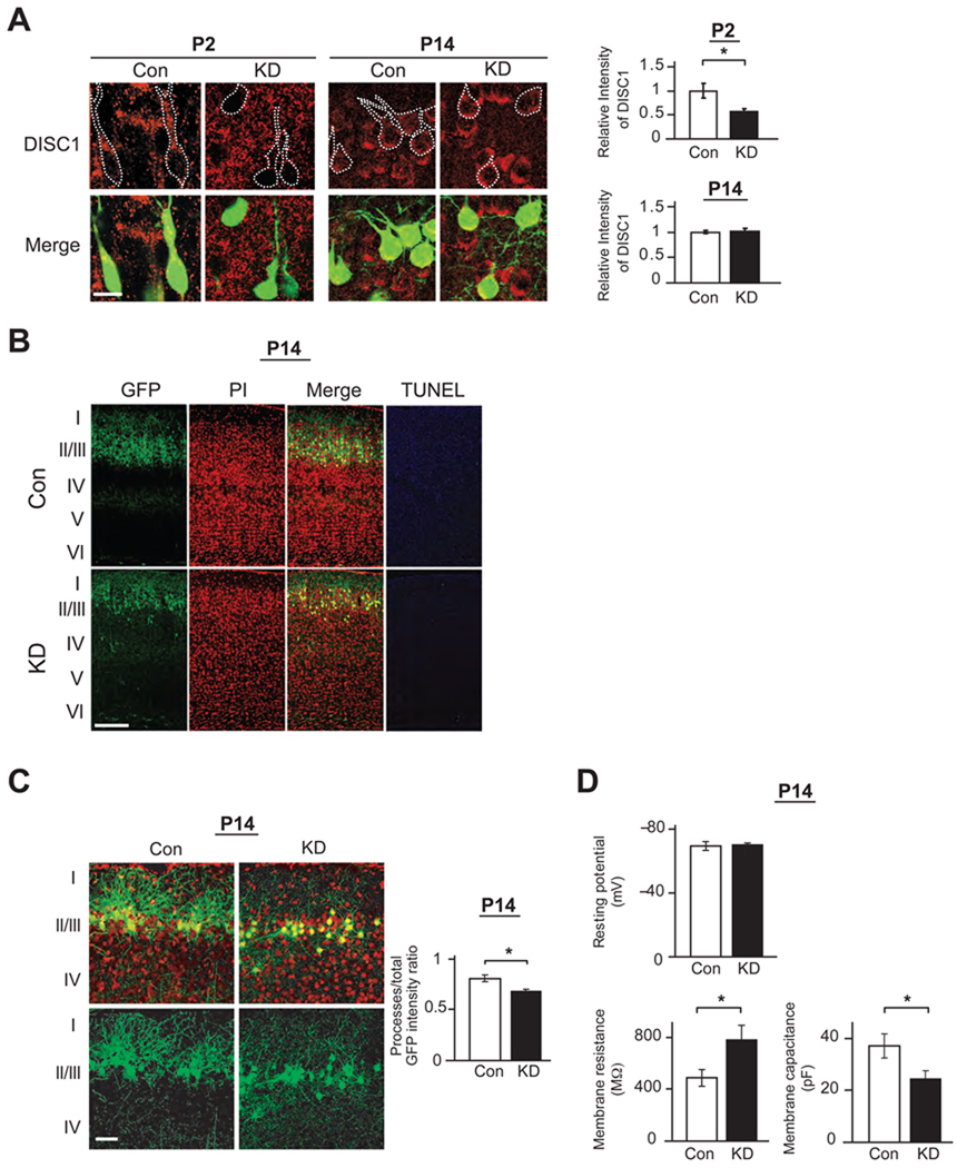
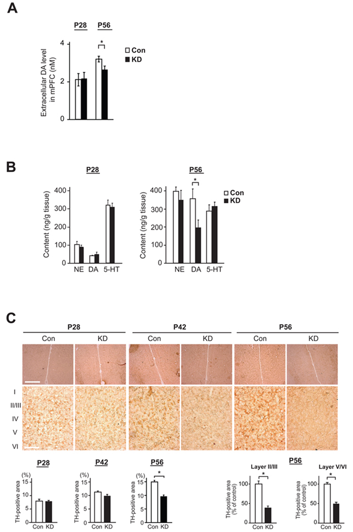
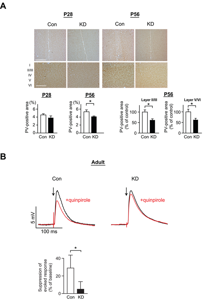
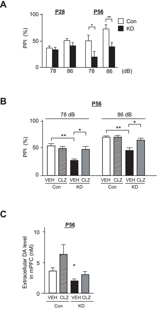
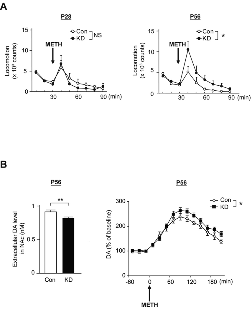
Adult brain function and behavior are influenced by neuronal network formation during development. Genetic susceptibility factors for adult psychiatric illnesses, such as Neuregulin-1 and Disrupted-in-Schizophrenia-1 (DISC1), influence adult high brain functions, including cognition and information processing. These factors have roles during neurodevelopment and are likely to cooperate, forming pathways or "signalosomes." Here we report the potential to generate an animal model via in utero gene transfer in order to address an important question of how nonlethal deficits in early development may affect postnatal brain maturation and high brain functions in adulthood, which are impaired in various psychiatric illnesses such as schizophrenia. We show that transient knockdown of DISC1 in the pre- and perinatal stages, specifically in a lineage of pyramidal neurons mainly in the prefrontal cortex, leads to selective abnormalities in postnatal mesocortical dopaminergic maturation and behavioral abnormalities associated with disturbed cortical neurocircuitry after puberty.


Figures

Figure 1
Figure 1. Selective targeting of constructs to cells in a lineage of pyramidal neurons in the prefrontal cortex via in utero gene transfer
(A) Schematic representation of bilateral in utero injection of constructs followed by their incorporation by electroporation into progenitor cells in the ventricular zone at embryonic day 14 (E14). Migrating cells with GFP are visualized at E18 after injection of a GFP expression construct. (B) Representative coronal and sagittal images of brains with GFP expression at P56 after injection of a GFP expression construct at E14. Blue, nucleus. Scale bar, 1 mm. (C) GFP-positive neurons in layers II/III at postnatal day 56 (P56) in mPFC. Most cells with GFP expression are CaMKII-positive pyramidal neurons (red in left panels), but not GABA (red in middle panels) or GFAP-positive (red in right panels) at P56. Scale bar, 100 µm.
Figure 2
Figure 2. Mice with knockdown of DISC1 in pyramidal neurons of the prefrontal cortex during early development via in utero gene transfer display dendritic abnormalities at P14
(A) Suppression of DISC1 immunoreactivity (red) in GFP-positive neurons is observed at P2, but not at P14, when shRNA to DISC1 is introduced [DISC1 knockdown (KD)] at E14 (*p<0.05). Total of 30 GFP-positive cells from cortical slices of 3 DISC1 KD mice and those of 3 control mice with scrambled/control shRNA were analyzed and compared. Scale bar, 10 µm. (B) GFP-positive neurons with RNAi at layers II/III at P14 after injection at E14. KD mice display dendritic pathology without signs of apoptosis (TUNEL), consistent with our previous publication (Kamiya et al., 2005). Red, nucleus. Scale bar, 100 µm. (C) Reduction of GFP fluorescence intensity in dendrites relative to total GFP fluorescence intensity in mPFC of KD mice at P14, compared to those of Con mice (*p<0.05, N=5 per condition), suggesting impaired dendritic formation in KD mice. Red, nucleus. Scale bar, 50 µm. (D) Electrophysiological characteristics of pyramidal neurons with knockdown of DISC1 (majority of green cells) at layer II/III in mPFC on P14. Membrane resistance at −80 mV and membrane capacitance of GFP-positive neurons in DISC1 KD mice are significantly different compared with those in Con mice (*p<0.05), whereas there is no difference in resting potential in these two groups (N=5 per condition).
Figure 3
Figure 3. Disturbance in postnatal maturation of mesocortical dopaminergic projections to the medial prefrontal cortex (mPFC) in DISC1 knockdown (DISC1 KD) mice
(A) Basal levels of extracellular dopamine (DA) in mPFC were analyzed by in vivo microdialysis, and decrease in DISC1 KD mice compared with that in Con mice was detected at P56, but not at P28 (*p<0.05, N=6 per condition). (B) Monoamine content in the frontal cortex (FC) measured by HPLC. Level of DA in FC is decreased in DISC1 KD mice compared with that in Con mice at P56, but not at P28 (*p<0.05, N=7 per condition). No difference in the levels of NE and 5-HT is observed at these two time points. (C) Immunostaining of tyrosine hydroxylase (TH) in mPFC, including prelimbic and infralimbic cortex (upper panels, low magnification; bottom panels, high magnification). TH level in mPFC is relatively decreased in KD mice compared to Con mice at P56 (*p<0.05, N=6 per condition), in both Layers II/III and V/VI, whereas there is no difference between KD and Con at P28 and P42. Scale bars in low magnification pictures, 200 µm; in high magnification pictures, 500 µm.
Figure 4
Figure 4. Disturbances of interneurons and pyramidal neurons in PFC of DISC1 KD mice after puberty
(A) Immunostaining of parvalbumin (PV) in the mPFC (upper panels, low magnification; bottom panels, high magnification). Immunoreactivity of PV is quantified in each condition (lower graphs). The expression levels of PV in the mPFC (both layers II/III and V/VI) are decreased in KD mice compared with Con mice at P56, but not P28 (*p<0.05, N=6 per condition). Scale bars in low magnification pictures, 200 µm; in high magnification pictures, 500 µm. (B) Electrophysiological responses of PFC pyramidal neurons to electrical stimulation recorded using the whole-cell patch-clamp technique in acute slices from young adult male mice. Overlay of membrane potential responses evoked with electrical stimulation of cortico-cortical fibers before (black trace) and during (red trace) bath application of the D2 dopamine agonist quinpirole (5 µM) in KD and Con mice. Arrows indicate times of single pulse stimulation. Quinpirole attenuation of evoked excitatory postsynaptic potentials (EPSPs) is reduced in KD mice (*p<0.05, N=5 per condition).
Figure 5
Figure 5. Attenuation of prepulse inhibition deficits by treatment with clozapine in DISC1 KD mice after puberty
(A) Performance in prepulse inhibition (PPI) at P28 and P56. Impairment of PPI is observed in DISC1 KD mice at P56, but not P28 (*p<0.05, **p<0.01, N=12 per condition). (B) Effect of clozapine (CLZ) on the impairment of PPI in KD mice at P56. Treatment with CLZ (3 mg/kg, i.p.) ameliorates the impairment of PPI in KD mice at P56 (**p<0.01) (N=16 per condition). VEH, vehicle. (C) Effect of CLZ on extracellular DA levels in mPFC by in vivo microdialysis. Administration of CLZ (3 mg/kg, i.p.) elevates extracellular DA levels in mPFC in both Con and KD mice at P56 (N=4 per condition).
Figure 6
Figure 6. Hypersensitivity to psychostimulant, methamphetamine, in DISC1 KD mice after puberty
(A) Methamphetamine (METH: 1 mg/kg, s.c.)-induced hyperactivity is augmented in DISC1 KD mice compared with Con mice, at P56 but not P28 (*p<0.05, N=6–10 per condition). (B) The extent of increase after METH challenge in levels of extracellular DA relative to those at the baseline is augmented in the nucleus accumbens (NAc) of DISC1 KD mice compared with Con mice, at P56 (*p<0.05, N=8 per condition) (right); whereas mild but significant reduction of basal levels of extracellular DA levels is observed in KD mice (*p<0.05, N=8 per condition) (left).

Comment in

References

    1. Akbarian S, Kim JJ, Potkin SG, Hagman JO, Tafazzoli A, Bunney WE, Jr, Jones EG. Gene expression for glutamic acid decarboxylase is reduced without loss of neurons in prefrontal cortex of schizophrenics. Arch Gen Psychiatry. 1995;52:258–266. - PubMed
    1. Akil M, Pierri JN, Whitehead RE, Edgar CL, Mohila C, Sampson AR, Lewis DA. Lamina-specific alterations in the dopamine innervation of the prefrontal cortex in schizophrenic subjects. Am J Psychiatry. 1999;156:1580–1589. - PubMed
    1. Anderson SA, Eisenstat DD, Shi L, Rubenstein JL. Interneuron migration from basal forebrain to neocortex: dependence on Dlx genes. Science. 1997;278:474–476. - PubMed
    1. Arguello PA, Gogos JA. Modeling madness in mice: one piece at a time. Neuron. 2006;52:179–196. - PubMed
    1. Benes FM, Berretta S. GABAergic interneurons: implications for understanding schizophrenia and bipolar disorder. Neuropsychopharmacology. 2001;25:1–27. - PubMed

Publication types

MeSH terms

LinkOut - more resources