. 2010 Feb 25;65(4):490-502.
doi: 10.1016/j.neuron.2010.02.004.

A glial signal consisting of gliomedin and NrCAM clusters axonal Na+ channels during the formation of nodes of Ranvier

Affiliations

A glial signal consisting of gliomedin and NrCAM clusters axonal Na+ channels during the formation of nodes of Ranvier

Konstantin Feinberg et al. Neuron. .

Abstract

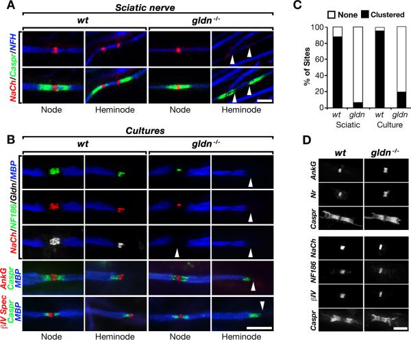
Saltatory conduction requires high-density accumulation of Na(+) channels at the nodes of Ranvier. Nodal Na(+) channel clustering in the peripheral nervous system is regulated by myelinating Schwann cells through unknown mechanisms. During development, Na(+) channels are first clustered at heminodes that border each myelin segment, and later in the mature nodes that are formed by the fusion of two heminodes. Here, we show that initial clustering of Na(+) channels at heminodes requires glial NrCAM and gliomedin, as well as their axonal receptor neurofascin 186 (NF186). We further demonstrate that heminodal clustering coincides with a second, paranodal junction (PNJ)-dependent mechanism that allows Na(+) channels to accumulate at mature nodes by restricting their distribution between two growing myelin internodes. We propose that Schwann cells assemble the nodes of Ranvier by capturing Na(+) channels at heminodes and by constraining their distribution to the nodal gap. Together, these two cooperating mechanisms ensure fast and efficient conduction in myelinated nerves.


Figures

Fig. 1
Fig. 1. Mice lacking gliomedin exhibit disorganized Schwann cell microvilli
A. Generation of gldn−/− mice. Schematic map of a genomic DNA fragment containing exons 4–10, the targeting construct used and the resulting allele lacking exon 9-10, which encode for the olfactomedin domain of gliomedin. B, BglII; S, SalI. B. Southern blot analysis of genomic DNA of wild type (wt) or knockout (ko) mice. The location of the probes and the expected fragments are labeled in the first panel. C. RT-PCR analysis of DRG mRNA using primer pairs present in exons 2-6 (Ex2-6), 6-9 (Ex6-9) and exon 10 (Ex10) revealed the absence of gliomedin transcript in homozygote mice. Primers for actin were used as controls. D. Western blot analysis of sciatic nerve lysates using antibodies to gliomedin (Gldn) or MBP as indicated. The location of molecular mass markers is indicated on the right in kDa. E. Gliomedin is absent from nodes in sciatic nerves of gldn−/− mice. Teased sciatic nerves of wild type (wt) and gldn−/− mice were labeled using antibodies to gliomedin (Gldn) and Caspr. Inset shows staining of nerves using a different antibody to gliomedin. Arrowheads mark the location of nodes. F. Gliomedin null mice exhibit normal PNS myelin. Left panels, immunolabeling of adult sciatic nerves isolated from wt and gldn−/− using antibodies to MAG and Caspr. Electron microscopy images of sciatic nerves cross sections are shown on the right panels. G. Schwann cell microvilli are disorganized in the absence of gliomedin. Electron microscopy images of sciatic nerves sectioned at nodes of P30 (three left panels) and P12 (right panel) mice. In gldn−/− mutant nerves, the microvilli are frequently directed in parallel to the axon and do not contact the axolemma. Scale bars: E, 10μm; F, 10μm (immunolabeling) and 0.25μm (EM); G, 0.5μm.
Fig. 2
Fig. 2. Gliomedin clusters Na+ channels at heminodes
A. Na+ channels are not clustered at heminodes but are present at nodes in gldn−/− mice. Sciatic nerve fibers of P6 wild type (wt) and gliomedin deficient (gldn−/−) mice labeled using antibodies to Na+ channels (NaCh), neurofilament (NFH) and Caspr. For each genotype nodes and heminodes are shown. Arrowheads mark the location of heminodes lacking Na+ channels. B. Myelinated Schwann/DRG neurons cultures isolated from wt and gldn−/− mice were labeled with the indicated antibodies. Na+ channels, NF186, ankyrin G (AnkG) and βIV-spectrin (βIV) are all absent from heminodes in cultures lacking gliomedin (arrowheads). An antibody to MBP was used to label myelin internodes. C. Quantification of the appearance of heminodal Na+ channels in sciatic nerve fibers (Sciatic) or myelinated cultures (SC/DRG) isolated from wt and gldn−/− mice; n=150 sites for sciatic nerves, n=300 for myelinating cultures (p < 0.001). D. Nodes of adult gldn−/− mice contain all components of the nodal complex. Teased sciatic nerves were labeled with antibodies to Na+ channels (NaCh), the axonodal CAMs (NrCAM and NF186), and to the nodal cytoskeletal proteins (βIV Spectrin and ankyrin G). An antibody to Caspr was used to mark the paranodal junctions bordering the nodes. Scale bars: A, 5μm; B, 10μm; D, 5μm. See also Figure S1.
Fig. 3
Fig. 3. Na+ channel clustering at heminodes requires both NrCAM and NF186
A. P6 sciatic nerve fibers of nrcam null mice (nrcam−/−) were labeled using antibodies to Na+ channels (NaCh), neurofilament (NFH) and Caspr. Arrowheads indicate the absence of Na+ channel clustering at heminodes. B. Myelinated Schwann/DRG neurons cultures isolated from wt and nrcam−/− mice were labeled with the indicated antibodies. Na+ channels, NF186 and gliomedin are absent from heminodes (arrowheads), but are present at mature nodes in nrcam−/− cultures. An antibody to MBP was used to label myelin internodes. C. Na+ channels are not clustered at heminodes in myelinated axons lacking NF186. Wild type Schwann cells were allowed to myelinate wild type (wt), or neurofascin null DRG neurons (wtSC;nfasc−/−N). Cultures were immunolabeled using antibodies to MBP, Caspr, Na+ channels (NaCh) and NF186. In wtSC;nfasc−/−N cultures (which lacks NF186), Caspr is present at the paranodal junction and Na+ channels accumulate at nodes, but not in heminodes. D. Appearance of heminodal Na+ channel clusters in nrcam−/− sciatic nerve, nrcam−/− myelinated cultures, or cultures containing wt Schwann cells and nfasc−/− neurons (wtSC;nfasc−/−N, i.e., nf186−/−); n=150 sites for sciatic nerves, n=300 for myelinating cultures (p < 0.001). E. Percentage of mature nodes containing (NaCh), or lacking (none), Na+ channels in wild type (wt) myelinated cultures, cultures of wild type Schwann cells and neurofascin null neurons (wtSC;nfasc−/−N, i.e., nf186−/−), or cultures of wild type Schwann cells and neurons isolated from double null mice lacking both NrCAM and neurofascin (wtSC;nrcam−/−/nfasc−/−N); n=200 (p < 0.001). F. Na+ channels accumulate in mature nodes in the absence of the axonodal CAMs. Myelinating co-cultures containing wild type Schwann cells and DRG neurons isolated from mice lacking NrCAM (wtSC;nr−/−), or both NrCAM and NF186 (wtSC;nf−/−/nr−/−N), were labeled using the indicated antibodies. Arrowhead marks the presence of Na+ channels at nodes in the absence of axonal NF186 and NrCAM. Scale bars: 5μm. See also Figure S2.
Fig. 4
Fig. 4. Gliomedin-dependent Na+ channel clustering is mediated by NF186
A. Gliomedin binds to both NF186 and NrCAM. Binding of soluble Fc-fusion protein containing the extracellular domain of gliomedin (Gldn-Fc) to DRG neurons isolated from wild type (wt), nrcam−/−, nfasc−/− or double nrcam−/−/nfasc−/− mutant mice. The presence of neurons was monitored by immunolabeling for βIII-tubulin (shown at lower magnification in insets). Note the absence of Gldn-Fc binding to DRG neurons lacking both NF186 and NrCAM. B. Na+ channel clustering requires NF186 but not NrCAM. Gldn-Fc was mixed with Cy3-conjugated secondary antibody to human Fc, and incubated with DRG neurons for 48 hours before fixing. Binding of Gldn-Fc is shown in the upper panels, along with immunofluorescence labeling for Na+ channels (NaCh) and the merged images. Arrowheads indicate the absence of Na+ channel clusters in nfasc−/− neurons. C. NrCAM clustered by gliomedin lacks its intracellular domain. DRG neurons were incubated with Gldn-Fc as described above and then fixed and immunolabeled using an antibody to neurofilament (NFH) together with antibodies that recognize the extracellular (NrCAM-ECD), or intracellular (NrCAM-Cyto) domain of NrCAM. Arrowhead depicts the absence of the intracellular domain of NrCAM in the cluster. Scale bars, 10μm.
Fig. 5
Fig. 5. NrCAM and gliomedin provide a glial signal for Na+ channel clustering
A. NrCAM is expressed by Schwann cells. RT-PCR analysis using primers for nrcam, gldn or actin on mRNA isolated from brain, mixed DRG/SC culture (DRG), and isolated mouse (mSC) or rat (rSC) Schwann cells. NrCAM cDNA (NrCAM) and reaction mix without template (none) were used as positive and negative controls, respectively. B. Schwann cells express a transmembrane and a secreted form of NrCAM. Cell lysates of Schwann cells and control 3T3 fibroblasts, or the growth medium of cultured Schwann cells were subjected to immunoprecipitation and Western blot analysis using an antibody to the extracellular domain of NrCAM. The location of molecular mass markers is indicated on the right in kDa. C. Neuronal expression of NrCAM is not sufficient for heminodal Na+ channel clustering. Myelinating cultures were prepared using wild type Schwann cells and DRG neurons (wt), or nrcam−/− Schwann cells and wild type neurons (nrcam−/− SC;wt N). In the absence of glial NrCAM (i.e., in nrcam−/− SC;wt N cultures), NF186, Na+ channels (NaCh) and axonal NrCAM accumulated in mature nodes, but did not clustered in heminodes (arrowheads). D. Glial NrCAM induces heminodal clustering of Na+ channels. Myelinating nrcam−/− cultures, or cultures prepared using nrcam−/− neurons and wild type Schwann cells (wt SC;nrcam−/− N) were immunolabeled using the indicated antibodies. In contrast to nrcam−/− cultures (arrowheads), in wt SC;nrcam−/− N cultures, glial NrCAM clustered at heminodes together with NF186, Na+ channels (NaCh) and gliomedin (Gldn). Scale bars, 5μm. See also Figure S3.
Fig. 6
Fig. 6. The PNJ restrict the area occupied by Na+ channels between two myelin segments
A. Developmental stages of node formation. Myelinated Schwann cell/DRG neurons cultures obtained from wild type (wt), gldn−/− and nrcam−/− mice, or myelinating co-cultures consisting of wild type Schwann cells and DRG neurons isolated from nfasc−/− mice (nf186−/−) were labeled using antibodies to Na+ channels (NaCh), NF186 and MBP. Merged images of three different stages of the developing nodal gaps found in the same culture. Arrowheads indicate both heminodes and nodes. In the absence of either gliomedin, NrCAM or NF186, Na+ channels are initially scattered between two distant myelin segments and then accumulate at mature nodes. Note that in gldn−/− and nrcam−/− cultures, NF186 is missing from the area occupied by scattered Na+ channels, but emerges later in mature nodes. B. Na+ channels invade the internodal region below the compact myelin in the absence of PNJ. Myelinated cultures of the indicated genotypes were immunolabeled using antibodies to Na+ channels (NaCh), Caspr (to mark the PNJ) and MBP (to label compact myelin). Images show examples of nodal gaps that are bordered by the PNJ only on one side. The location of the paranodes (p), nodes (n) and internodes (i) is marked by vertical dashed lines. Arrowheads marks heminodal clustering of Na+ channels in wild type cultures. C. Scheme illustrating the distribution of Na+ channels (red) and Caspr (green) detected in panel B. D. Heminodes are formed in the absence of PNJ. Na+ channels co-cluster with gliomedin at heminodes in myelinated cultures of Schwann cell/DRG neurons isolated from caspr−/− mice. Scale bars: A-D, 5μm. See also Figure S4.
Fig. 7
Fig. 7. Assembly of the nodes of Ranvier requires axoglial contacts at nodes and paranodes
A. A reduction in Na+ channel clustering in gldn−/−/caspr−/− and nrcam−/−/caspr−/− mice. Longitudinal sections of P6 sciatic nerves isolated from the indicated genotypes, immunolabeled with antibodies to neurofilament (NFH) and Na+ channels (NaCh). Higher magnification of representative nodal sites lacking Na+ channels (arrowheads) in sciatic nerves of the double mutants is shown in the lower panels. B. Amount of Na+ channels clusters per field of view (FOV); error bars, SD of n=15-20 fields for each genotype (**p < 0.001). C. Examples of nodal sites that formed in gldn−/−/caspr−/− and nrcam−/−/caspr−/− mice. P6 sciatic nerves isolated from wild type (wt), gldn−/−/caspr−/− and nrcam−/−/caspr−/− mice, were immunolabeled using antibodies to neurofilament (NFH), Na+ channels (NaCh) and βIV spectrin (βIV). Higher magnification of the boxed area is shown below each panel. D. Quantification of the areas occupied by Na+ channels co-localized with βIV spectrin in the different genotypes; error bars, SD of n=100 sites for each genotype (**p < 0.001). E-F. Nerve conduction velocity (NCV) is reduced in double mutant mice lacking Caspr and either gliomedin or NrCAM. Compound action potentials were recorded from sciatic nerves of P14 (E) or P7 (F) animals; error bars, SEM of n=7 mice for each genotype (**p < 0.005). Scale bars: A, 10μm; C, 5μm. See also Figure S5.
Fig. 8
Fig. 8. Two distinct axoglial adhesion systems assemble PNS nodes of Ranvier
A. Na+ channels (red circle) are trapped at heminodes that are contacted by Schwann cell microvilli (MV; orange). Axon-glia interaction at this site is mediated by binding of gliomedin and glial NrCAM to axonal NF186 (D). A transmembrane and secreted forms of glial NrCAM trap gliomedin on Schwann cell microvilli and enhances its binding to NF186. In the absence of either gliomedin or glial NrCAM, Na+ channels fail to cluster at heminodes. Binding of gliomedin to axonal NrCAM, which lack its cytoplasmic domain does not result in Na+ channel clustering. B. The distribution of Na+ channels is restricted between two forming myelin segments by the PNJ (blue). Three CAMs, NF155 present at the paranodal loops (green) and axonal complex of Caspr and contactin mediate axon-glia interaction and the formation of the PNJ (E). C. These two cooperating mechanisms provide reciprocal backup systems and ensure that Na+ channels are found at high density at the nodes.

Similar articles

Cited by

References

    1. Arroyo EJ, Sirkowski EE, Chitale R, Scherer SS. Acute demyelination disrupts the molecular organization of peripheral nervous system nodes. J Comp Neurol. 2004;479:424–434. - PubMed
    1. Berghs S, Aggujaro D, Dirkx R, Jr., Maksimova E, Stabach P, Hermel JM, Zhang JP, Philbrick W, Slepnev V, Ort T, Solimena M. betaIV spectrin, a new spectrin localized at axon initial segments and nodes of ranvier in the central and peripheral nervous system. J Cell Biol. 2000;151:985–1002. - PMC - PubMed
    1. Berthold CH, Rydmark M. Electron microscopic serial section analysis of nodes of Ranvier in lumbosacral spinal roots of the cat: ultrastructural organization of nodal compartments in fibres of different sizes. J Neurocytol. 1983;12:475–505. - PubMed
    1. Bhat MA, Rios JC, Lu Y, Garcia-Fresco GP, Ching W, St Martin M, Li J, Einheber S, Chesler M, Rosenbluth J, et al. Axon-glia interactions and the domain organization of myelinated axons requires neurexin IV/Caspr/Paranodin. Neuron. 2001;30:369–383. - PubMed
    1. Boyle ME, Berglund EO, Murai KK, Weber L, Peles E, Ranscht B. Contactin orchestrates assembly of the septate-like junctions at the paranode in myelinated peripheral nerve. Neuron. 2001;30:385–397. - PubMed

Publication types

MeSH terms