. 2010 Feb 26;37(4):469-80.
doi: 10.1016/j.molcel.2010.01.020.

A role for Gcn5 in replication-coupled nucleosome assembly

Affiliations

A role for Gcn5 in replication-coupled nucleosome assembly

Rebecca J Burgess et al. Mol Cell. .

Abstract

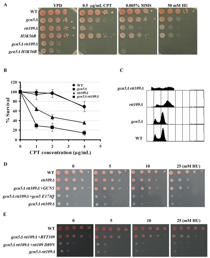
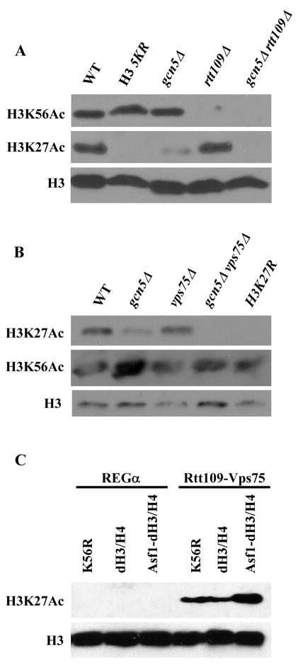
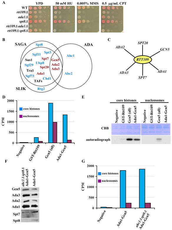
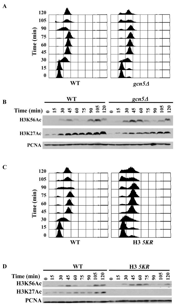
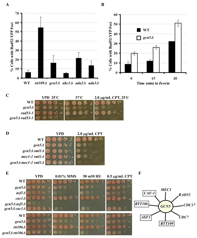
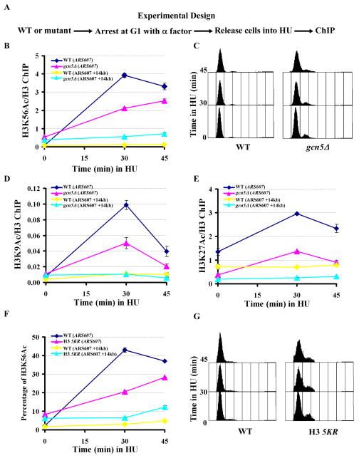
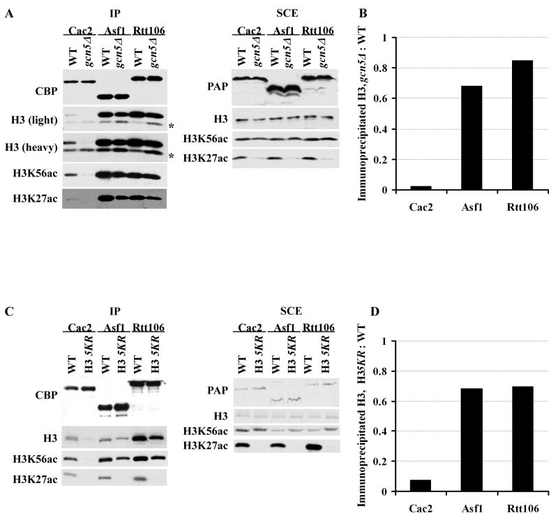
Acetylation of lysine residues at the H3 N terminus is proposed to function in replication-coupled (RC) nucleosome assembly, a process critical for the inheritance of epigenetic information and maintenance of genome stability. However, the role of H3 N-terminal lysine acetylation and the corresponding lysine acetyltransferase (KAT) in RC nucleosome assembly are not known. Here we show that Gcn5, a KAT that functions in transcription, works in parallel with Rtt109, the H3 lysine 56 KAT, to promote RC nucleosome assembly. Cells lacking both Gcn5 and Rtt109 are highly sensitive to DNA damaging agents. Moreover, cells lacking GCN5 or those that express N-terminal H3 mutants are compromised for deposition of new H3 onto replicating DNA and also show reduced binding of H3 to CAF-1, a histone chaperone involved in RC nucleosome assembly. These results demonstrate that Gcn5 regulates RC nucleosome assembly, in part, by promoting H3 association with CAF-1 via H3 acetylation.


Figures

Figure 1
Figure 1
Gcn5 and H3 lysine 56 acetylation function in parallel in growth and response to DNA damaging agents. Data presented in Table S1 indicate the presence of synthetic genetic interactions between the HAT, GCN5, and ASF1, a regulator of H3K56Ac. (A) The gcn5Δ mutant exhibits synthetic phenotypes with rtt109Δ and H3K56R mutants. Ten fold serial dilutions of wild-type (WT) or mutant yeast cells with relevant genotype indicated were spotted onto normal growth media, YPD, or media containing the indicated concentration of the DNA damaging agents hydroxyurea (HU), methyl methane sulfonate (MMS), or camptothecin (CPT). Full data presented in Figure S1. (B) The gcn5Δ rtt109Δ double mutant cells are more sensitive to CPT than either single mutant. Yeast cells were treated with the indicated concentration of CPT for 2 hours, and the percentage of surviving cells was reported. (C) FACS analysis of the DNA content of unsynchronized yeast cells. (D–E) The catalytic activity of Gcn5 (D) and Rtt109 (E) is required for cell growth and sensitivity towards DNA damaging agents. Mutant cells transformed with either plasmid for wild-type GCN5, gcn5 E173Q, or empty vector (gcn5Δ rtt109Δ) were spotted onto media containing different concentrations of HU (D). Similar experiments were also performed for wild-type RTT109 or the rtt109 D89N (E).
Figure 2
Figure 2
Both Gcn5 and Rtt109 acetylate H3 lysine 27 (H3K27Ac). (A) Cells lacking both GCN5 and RTT109 result in a significant loss of H3K27Ac. The H3 5KR mutant contains mutations at five lysine residues of the N-terminus of H3 (9, 14, 18, 23 and 27). (B) Vps75 is essential for H3K27Ac in the absence of Gcn5. Western blot analysis of whole cell extracts showing loss of H3K27Ac in gcn5Δ vps75Δ cells. H3K27R: lysine 27 of H3 was mutated to arginine. (C) The Rtt109-Vps75 complex acetylates H3K27 in vitro. HAT assays were performed using recombinant histone H3-H4 in the presence or absence of the histone chaperone Asf1. Western blot was performed using antibodies against H3 or H3K27Ac.
Figure 3
Figure 3
A Gcn5-containing complex functions in parallel with Rtt109 in a common process. (A) Mutations in ADA1 but not SPT8 phenocopy the gcn5Δ mutant in the absence of Rtt109. (B) Venn diagram depicting the subunits of the SAGA, SLIK, and ADA complex. Genes in black represent those that were not tested, those in blue are genes that did not show synergistic phenotypes, and in red, are genes that exhibited synthetic phenotypes when combined with deletion of RTT109. Complete genetic data is in Figure S2. (C) Web depicting genetic interactions with RTT109. (D and E) The proteins that co-purified with Ada1 and Gcn5 (Ada1-Gcn5) have HAT activities different from those purified with Gcn5-TAP (Gcn5-all). Core histones and mono-nucleosomes were used as substrates for the HAT assays and incorporated 3H-CoA acetate was quantified (D). The HAT assay samples were also resolved on SDS-PAGE and revealed by Coomassie Brilliant Blue staining (CBB) (E, top) or by fluorography (E, bottom). (F) Western blot analysis of indicated components of the Ada1-Gcn5 protein complex purified from wild-type (Ada1-Gcn5, Ada1-TAP Gcn5-Flag) cells or cells lacking Ahc1 and Spt8 (ahc1Δ spt8Δ). (G) The HAT activity of complexes purified from wild-type (Ada1-Gcn5) cells and ahc1Δ spt8Δ cells.
Figure 4
Figure 4
The gcn5Δ and H3 5KR mutants affect cell cycle progression and cell cycle dynamics of H3K56Ac. (A-B) The gcn5Δ cells exhibit a prolonged G2/M phase and persistence of high levels of H3K56Ac compared to wild-type cells. Every 15 minutes after release from G1, aliquots of cells were removed for analysis of DNA content (A) or analysis of H3K56Ac and H3K27Ac by Western blot (B). (C-D) Cells expressing the H3 5KR mutant showed a prolonged G2/M phase and persistence of a high level of H3K56Ac compared to wild-type cells. The experiments were performed as described in A and B.
Figure 5
Figure 5
GCN5 is involved in DNA replication-coupled nucleosome assembly. (A) Cells lacking GCN5, ADA2 or ADA3 exhibited a higher level of Rad52 foci compared to wild-type cells. The percentage of cells with Rad52-YFP foci was determined as described in Experimental Procedures. Data represent the mean percentage ± SEM. Mock experiments were also performed in which the genotype of each strain was unknown to the person who performed the experiment. (B) The gcn5Δ mutant cells exhibited higher levels of chromosome breaks than wild-type cells when challenged with a DNA damaging agent. Wild-type or gcn5Δ cells expressing Rad52-YFP were treated with zeocin for 0, 15, or 30 minutes and the percentage of cells containing Rad52 foci was determined. (C–D) The gcn5Δ mutation exhibited a synthetic genetic interaction with mutations in the checkpoint kinases RAD53 (C, rad53-1) and MEC1 (D, mec1-1). (E) The gcn5Δ mutant exhibited slow growth and increased DNA damage sensitivity with mutations in ASF1, CAC1, and RTT106. (F) A summary of genetic interactions observed for GCN5. Those in blue boxes indicate genes that are known to be involved in nucleosome assembly. Complete genetic data is presented in Figure S3.
Figure 6
Figure 6
Gcn5 and five lysine residues at the N-terminus of H3 are required for efficient deposition of newly-synthesized H3 onto replicating DNA. (A) A schematic diagram showing ChIP experimental design. Wild-type or mutant cells (gcn5Δ or H3 5KR) were arrested in G1 using α-factor and then released into HU. At different time points, samples were removed for FACS analysis and ChIP assays using antibodies against H3 or H3K56Ac. (B) The deposition of H3K56Ac onto replicating DNA is compromised in gcn5Δ mutant cells. The ChIP DNA was analyzed using primers that amplify the replication origin ARS607 or a fragment 14 kb away from ARS607 (ARS607+14 kb). The ratio of H3K56Ac ChIP signal over that of H3 was calculated. (C) FACS analysis of the DNA content of wild-type and gcn5Δ mutant cells used in B. (D–E) H3K9Ac (D) and H3K27Ac (E) were detected at replicating DNA. The experiment was performed as described in A and B except that antibodies against H3K9Ac or H3K27Ac were used. (F) Deposition of H3K56Ac was compromised in cells expressing the H3 5KR mutant cells. The experiment was performed as described in A and B except that the percentage of DNA that was precipitated by antibodies against H3K56Ac was calculated by analyzing both ChIP DNA and DNA from whole cells using real-time PCR. (G) FACS analysis of the DNA content of yeast strains used in experiments described in F. Each ChIP experiment was performed independently at least twice. The data represent mean ± SEM of three real-time PCR samples from one experiment. See also Figure S4.
Figure 7
Figure 7
Gcn5 and acetylation of five lysine residues of the H3 N-terminus regulate the binding of H3 with CAF-1. (A–B) The binding of H3 with CAF-1 is significantly reduced in gcn5Δ mutant cells. (A) Cac2-, Asf1- and Rtt106-TAP were purified from wild-type and gcn5Δ mutant cells, and co-purified proteins as well as proteins in soluble cell extracts (SCE) were detected by Western blot using antibodies against H3, H3K56ac, H3K27ac, calmodulin binding peptide (CBP) and IgG (PAP). * indicates non-specific band. (B) Quantification of the co-purified H3. The H3 intensity shown in A was quantified, and the ratio of the H3 that co-purified from each protein from the gcn5Δ cells over wild type cells was reported. (C–D) The association of H3 with CAF-1 is significantly reduced in cells expressing the H3 5KR mutant cells. The experiment was performed as described in A and quantification in D was performed as described in B.

Comment in

Similar articles

Cited by

References

    1. Ai X, Parthun MR. The nuclear Hat1p/Hat2p complex: a molecular link between type B histone acetyltransferases and chromatin assembly. Mol Cell. 2004;14:195–205. - PubMed
    1. Balasubramanian R, Pray-Grant MG, Selleck W, Grant PA, Tan S. Role of the Ada2 and Ada3 transcriptional coactivators in histone acetylation. J Biol Chem. 2002;277:7989–7995. - PubMed
    1. Benson LJ, Gu Y, Yakovleva T, Tong K, Barrows C, Strack CL, Cook RG, Mizzen CA, Annunziato AT. Modifications of H3 and H4 during chromatin replication, nucleosome assembly, and histone exchange. J Biol Chem. 2006;281:9287–9296. - PubMed
    1. Berndsen CE, Tsubota T, Lindner SE, Lee S, Holton JM, Kaufman PD, Keck JL, Denu JM. Molecular functions of the histone acetyltransferase chaperone complex Rtt109-Vps75. Nat Struct Mol Biol. 2008;15:948–956. - PMC - PubMed
    1. Candau R, Berger SL. Structural and functional analysis of yeast putative adaptors. Evidence for an adaptor complex in vivo. J Biol Chem. 1996;271:5237–5245. - PubMed

Publication types