. 2010 Feb 26;37(4):492-502.
doi: 10.1016/j.molcel.2010.01.021.

Hydroxyurea-stalled replication forks become progressively inactivated and require two different RAD51-mediated pathways for restart and repair

Affiliations

Hydroxyurea-stalled replication forks become progressively inactivated and require two different RAD51-mediated pathways for restart and repair

Eva Petermann et al. Mol Cell. .

Abstract

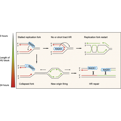
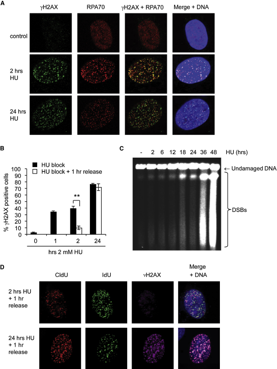
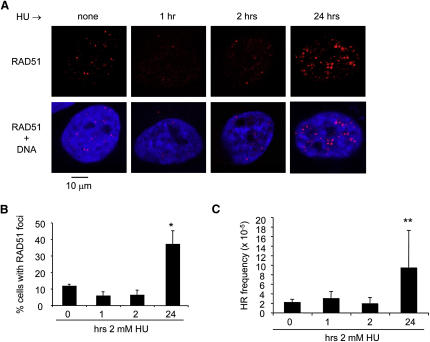
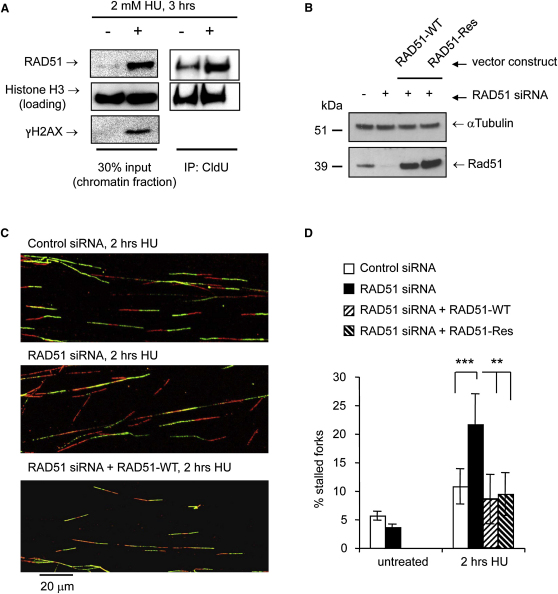
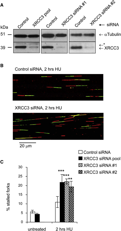
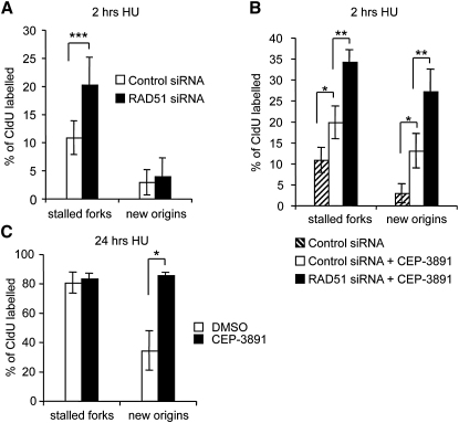
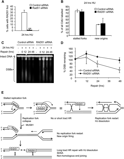
Faithful DNA replication is essential to all life. Hydroxyurea (HU) depletes the cells of dNTPs, which initially results in stalled replication forks that, after prolonged treatment, collapse into DSBs. Here, we report that stalled replication forks are efficiently restarted in a RAD51-dependent process that does not trigger homologous recombination (HR). The XRCC3 protein, which is required for RAD51 foci formation, is also required for replication restart of HU-stalled forks, suggesting that RAD51-mediated strand invasion supports fork restart. In contrast, replication forks collapsed by prolonged replication blocks do not restart, and global replication is rescued by new origin firing. We find that RAD51-dependent HR is triggered for repair of collapsed replication forks, without apparent restart. In conclusion, our data suggest that restart of stalled replication forks and HR repair of collapsed replication forks require two distinct RAD51-mediated pathways.


Figures

None
Graphical abstract
Figure 1
Figure 1
Replication Forks Restart after Short Replication Blocks but Become Inactivated during Long Replication Blocks (A) Labeling protocols for DNA fiber and replication foci analysis. U2OS cells were pulse-labeled with CldU, treated with 2 mM HU for the times indicated, and released into IdU. CldU was detected using a specific primary antibody and a secondary antibody in red. IdU was detected using specific primary antibody and a secondary antibody in green. (B) Representative images of replication tracks from U2OS cells after release from 1, 2, or 24 hr HU treatment. (C) Quantification of fork restart in U2OS cells after release from 1, 2, or 24 hr HU treatment. (D) Global replication restart in U2OS cells after release from 2 or 24 hr HU treatment. Cells were pulse-labeled as in (A), then fixed and immunostained for CldU (red) and IdU (green). DNA was counterstained with DAPI (blue). (E) Quantification of cells restarting global replication as in (D). Cells labeled with both CldU and IdU are shown as percentages of all cells labeled with CldU. (F) Quantification of cells newly entering S phase after release from 2 or 24 hr HU treatment. Cells labeled with only IdU are shown as percentages of total cells. The means and standard deviation (SD) (bars) of at least three independent experiments are shown. Values marked with asterisks are significantly different (Student's t test, p < 0.05, ∗∗p < 0.01, and ∗∗∗p < 0.001; see also Figure S1).
Figure 2
Figure 2
Replication Forks Accumulate DNA Damage during Long Replication Blocks (A) Colocalization of HU-induced γH2AX with RPA. Cells were left untreated or treated with 2 mM HU for 2 or 24 hr, fixed, and immunostained for γH2AX (green) and RPA70 (red). DNA was counterstained with DAPI (blue). (B) Quantification of U2OS cells displaying γH2AX immunostaining after 0, 1, 2, or 24 hr of treatment with 2 mM HU and 1 hr after release from 2 or 24 hr HU treatment. Cells containing more than 10 foci were scored as positive. The means and SD (bars) of three independent experiments are shown. Values marked with asterisks are significantly different (∗∗p < 0.01). (C) Pulsed-field gel electrophoresis to visualize DSB induction in U2OS cells treated with 2 mM HU for 2 to 48 hr. (D) Colocalization of γH2AX and stalled replication forks in U2OS cells. Cells were pulse-labeled with CldU for 20 min, treated with 2 mM HU for 2 or 24 hr, and released into IdU for 1 hr. Cells were immunostained for CldU (red), IdU (green), and γH2AX (far red), and DNA was counterstained with DAPI (blue). DNA was denatured with HCl to allow CldU/IdU detection.
Figure 3
Figure 3
Late Activation of Homologous Recombination during HU Block (A) HU-induced RAD51 foci in U2OS cells. Cells were left untreated or treated with 2 mM HU for 1, 2, or 24 hr, fixed, and immunostained for RAD51 (red). DNA was counterstained with DAPI (blue). (B) Quantification of HU-induced RAD51 foci in U2OS cells. Cells were treated with 2 mM HU for the times indicated and immunostained for RAD51. Cells containing >10 RAD51 foci were scored as positive. The means and range (bars) of two independent experiments are shown. (C) Recombination frequencies in SPD8 cells induced by HU. Cells were treated with 2 mM HU for the times indicated and recovered for 48 hr. HPRT+ revertants were quantified 7 days later. The means and SD (bars) of four to ten independent experiments are shown. Values marked with asterisks are significantly different from control (p < 0.05, ∗∗p < 0.01, and ∗∗∗p < 0.001; see also Figure S2).
Figure 4
Figure 4
Early Role of RAD51 in Promoting Replication Fork Restart (A) Localization of human RAD51 to replication forks by CldU coimmunoprecipitation (IP). U2OS cells were treated with 2 mM HU for 3 hr, released from HU, and pulse-labeled with CldU for 40 min. Cells were cross-linked, and the chromatin fraction was isolated (input) and subjected to IP using anti-CldU antibody (IP). Fractions were probed for RAD51, γH2AX, and Histone H3 (loading control). (B) Protein levels of RAD51 and α-Tubulin (loading control) in U2OS cells after 48 hr depletion with RAD51 or control siRNA. For re-expression of RAD51, cells were cotransfected with expression constructs encoding wild-type (RAD51-WT) or targeting-resistant (RAD51-Res) RAD51. (C) Representative images of replication tracks after restart from 2 hr HU treatment in control- and RAD51-depleted U2OS cells and RAD51-depleted cells re-expressing RAD51. Untreated values were only determined for control and RAD51 siRNA. (D) Quantification of fork restart in cells as in (C). Stalled replication forks are shown as percentage of all CldU labeled tracks. The means and SD (bars) of at least three independent experiments are shown. Values marked with asterisks are significantly different from control (Student's t test, ∗∗p < 0.01 and ∗∗∗p < 0.001).
Figure 5
Figure 5
XRCC3 Promotes the Early Restart of Stalled Replication Forks (A) Protein levels of XRCC3 and α-Tubulin (loading control) in U2OS cells after 48 hr depletion with control siRNA, pool of 4 XRCC3 siRNAs, or individual XRCC3 siRNAs #1 and #2. The asterisk denotes a nonspecific band. (B) Representative images of replication tracks after restart from 2 hr HU treatment in control- and XRCC3-depleted U2OS cells. (C) Quantification of fork restart in control- or XRCC3-depleted U2OS cells. The means and SD (bars) of at least three independent experiments are shown. Values marked with asterisks are significantly different from control (Student's t test, ∗∗p < 0.01 and ∗∗∗p < 0.001).
Figure 6
Figure 6
The S Phase Checkpoint Inhibits New Origin Firing Induced by Stalled Replication Forks (A) Quantification of fork restart and new origin firing in control- or RAD51-depleted U2OS cells after release from 2 hr HU treatment. Quantification of fork restart is as in Figure 4D. (B) Quantification of fork restart and new origin firing in U2OS cells after release from 2 hr HU treatment in presence or absence of Chk1 inhibitor CEP-3891 and RAD51 siRNA. CEP-3891 (500 nM) was present throughout HU treatment and during restart. (C) Quantification of fork restart and new origin firing in mock- or CEP-3891-treated U2OS cells after release from 24 hr HU treatment. CEP-3891 (500 nM) was added 1 hr before release from HU block and was present during restart. Replication structures are shown as percentage of all CldU-labeled tracks. The means and SD (bars) of three independent experiments are shown. Values marked with asterisks are significantly different from control (Student's t test, p < 0.05, ∗∗p < 0.01, and ∗∗∗p < 0.001; see also Figure S3).
Figure 7
Figure 7
After Long Replication Blocks, RAD51 Foci Do Not Promote Fork Restart but Are Required for DNA Damage Repair (A) Percentage of control- or RAD51-depleted U2OS cells containing more than 10 RAD51 foci after 24 hr treatment with 0.5 mM HU. The means and range (bars) of two independent experiments are shown. Values marked with asterisks are significantly different from control (p < 0.01). (B) Fork restart and new origin firing after release from 24 hr treatment with 2 mM HU in control- or RAD51-depleted U2OS cells. The means and SD (bars) of three independent experiments are shown. Replication structures are shown as percentage of all CldU-labeled tracks. (C) Pulsed-field gel electrophoresis to visualize DSB remaining in control- or RAD51-depleted U2OS cells after 0, 12 24, 36, and 48 hr release from 24 hr treatment with 2 mM HU. (D) Quantification of DSB remaining in control- or RAD51-depleted U2OS cells as in (C). The means and range (bars) of two to three independent experiments are shown. (E) Model of RAD51-mediated replication fork restart and repair. RAD51 may have a similar role as recA in E. coli, promoting the formation of a Holliday Junction intermediate (chicken foot). The DNA end may then assist to restart replication that would involve recombination over a small area (short tract). Holliday Junction dissolution by the BLM-Top3 complex would dissolve any remaining double Holliday Junctions (Wu and Hickson, 2003). New origin firing rescues replication of collapsed replication forks, which are then repaired by long tract HR involving double Holliday Junction dissolution, synthesis-dependent strand annealing (SDSA) or nonhomologous end joining (see also Figure S4).

Similar articles

Cited by

References

    1. Alabert C., Bianco J.N., Pasero P. Differential regulation of homologous recombination at DNA breaks and replication forks by the Mrc1 branch of the S-phase checkpoint. EMBO J. 2009;28:1131–1141. - PMC - PubMed
    1. Anglana M., Apiou F., Bensimon A., Debatisse M. Dynamics of DNA replication in mammalian somatic cells: nucleotide pool modulates origin choice and interorigin spacing. Cell. 2003;114:385–394. - PubMed
    1. Arnaudeau C., Lundin C., Helleday T. DNA double-strand breaks associated with replication forks are predominantly repaired by homologous recombination involving an exchange mechanism in mammalian cells. J. Mol. Biol. 2001;307:1235–1245. - PubMed
    1. Barlow J.H., Rothstein R. Rad52 recruitment is DNA replication independent and regulated by Cdc28 and the Mec1 kinase. EMBO J. 2009;28:1121–1130. - PMC - PubMed
    1. Baumann P., Benson F.E., West S.C. Human Rad51 protein promotes ATP-dependent homologous pairing and strand transfer reactions in vitro. Cell. 1996;87:757–766. - PubMed

Publication types