. 2010 Feb 26;37(4):529-40.
doi: 10.1016/j.molcel.2010.01.015.

The matrix peptide exporter HAF-1 signals a mitochondrial UPR by activating the transcription factor ZC376.7 in C. elegans

Affiliations

The matrix peptide exporter HAF-1 signals a mitochondrial UPR by activating the transcription factor ZC376.7 in C. elegans

Cole M Haynes et al. Mol Cell. .

Abstract

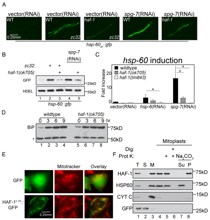
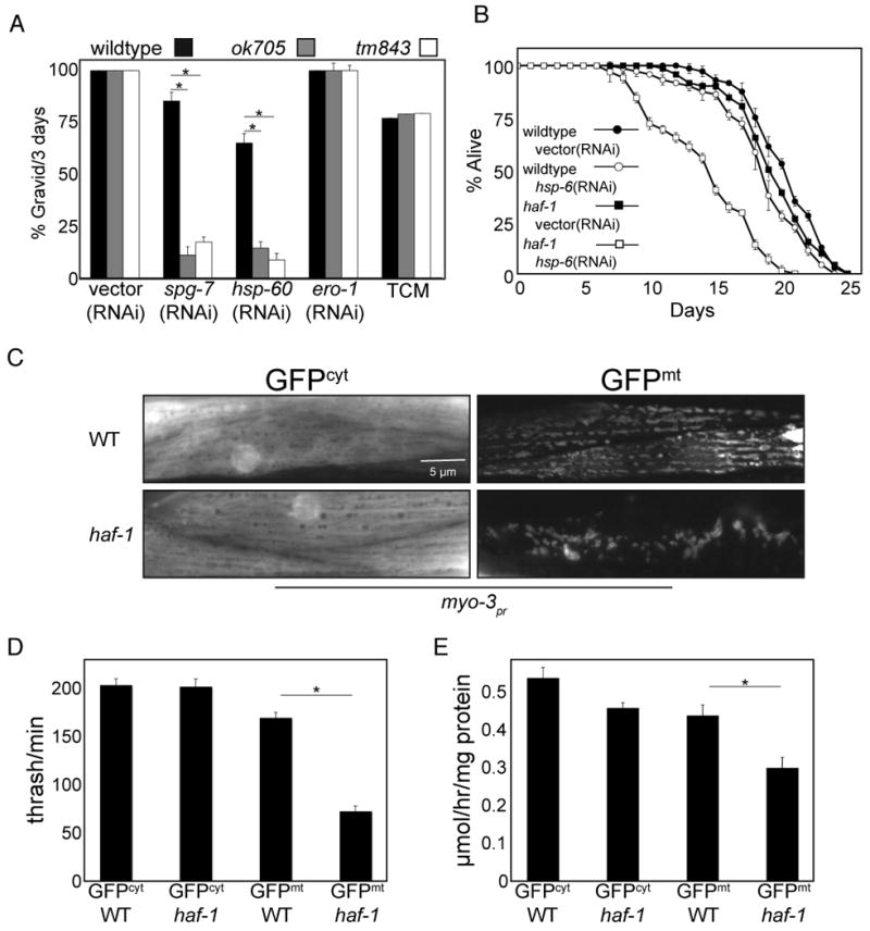
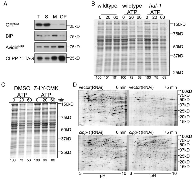
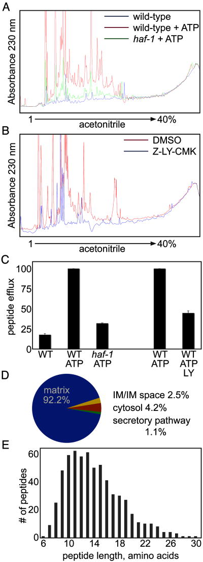
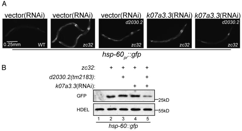
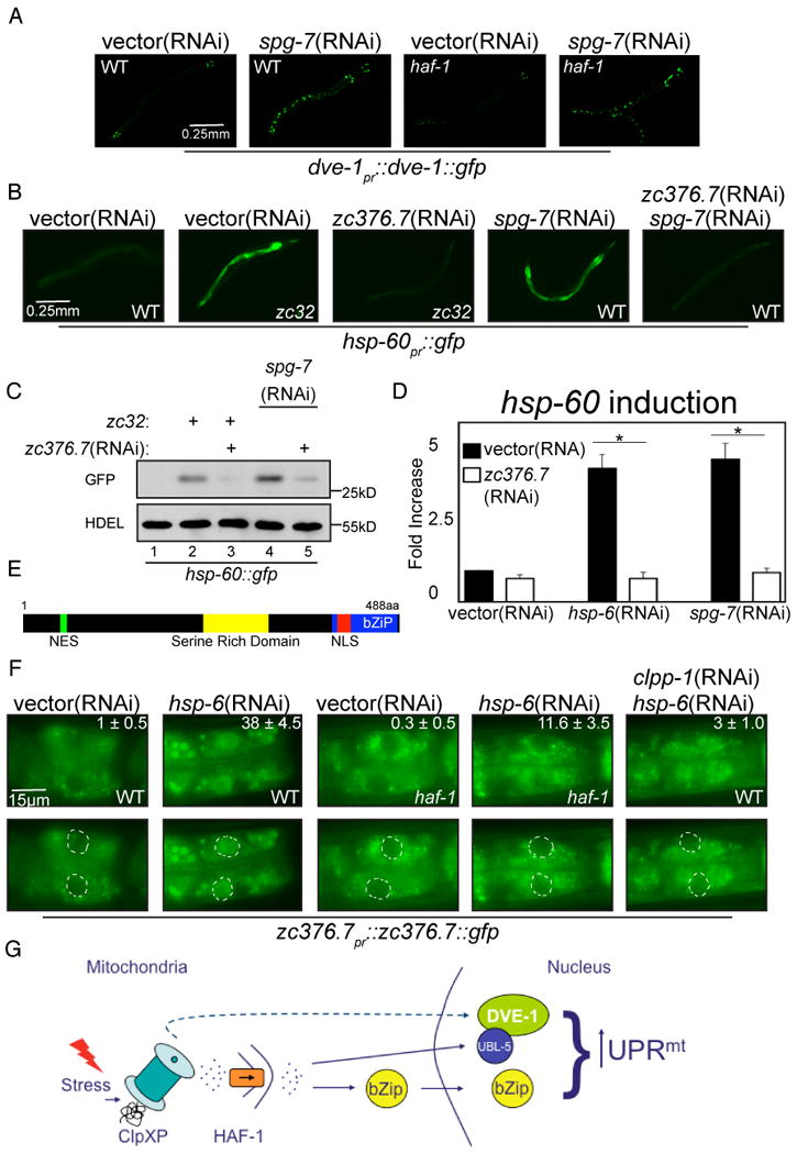
Genetic analyses previously implicated the matrix-localized protease ClpP in signaling the stress of protein misfolding in the mitochondrial matrix to activate nuclear-encoded mitochondrial chaperone genes in C. elegans (UPR(mt)). Here, we report that haf-1, a gene encoding a mitochondria-localized ATP-binding cassette protein, is required for signaling within the UPR(mt) and for coping with misfolded protein stress. Peptide efflux from isolated mitochondria was ATP dependent and required HAF-1 and the protease ClpP. Defective UPR(mt) signaling in the haf-1-deleted worms was associated with failure of the bZIP protein, ZC376.7, to localize to nuclei in worms with perturbed mitochondrial protein folding, whereas zc376.7(RNAi) strongly inhibited the UPR(mt). These observations suggest a simple model whereby perturbation of the protein-folding environment in the mitochondrial matrix promotes ClpP-mediated generation of peptides whose haf-1-dependent export from the matrix contributes to UPR(mt) signaling across the mitochondrial inner membrane.


Figures

Figure 1
Figure 1. Impaired UPRmt in haf-1 mutant animals
(A) Representative fluorescent photomicrographs of hsp-60pr∷gfp transgenic worms (reporting on the UPRmt) with a temperature sensitive mutation (zc32) that activates the UPRmt in an otherwise wildtype or haf-1(ok705) deleted background. Where indicated wildtype or haf-1(ok705) worms were raised on spg-7(RNAi) for 3 days to induce mitochondrial misfolded protein stress. (B) A corresponding immunoblot of GFP expressed by hsp-60pr∷gfp transgenic worms in whom mitochondrial unfolded protein stress was induced (as in “A”). The endogenous ∼55kDa ER protein, detected with an anti-HDEL monoclonal antibody (lower panel) serves as a loading control. (C) Quantitative analysis (by QRT-PCR) of endogenous hsp-60 mRNA in wildtype or two different haf-1 deleted strains (ok705 and tm843) subjected to mitochondrial unfolded protein stress by hsp-6(RNAi) or spg-7(RNAi). Displayed is the mean +/- SEM, n=3, *p<0.05. (D) Immunoblot of proteins reactive with an anti-HDEL monoclonal antibody in extracts of wildtype and haf-1 mutant worms in which ER stress was induced by exposure to elevated temperature (30°C) for the indicated time. The upper band, BiP (HSP-4), is a UPRER target gene whereas the lower invariant ∼55Kd band (*) serves as a loading control. (E) Fluorescent photomicrographs of Chinese Hamster Ovary (CHO) cells expressing GFP (upper panels) or GFP fused to amino acids 1-75 of HAF-1 (HAF-11-75∷GFP, lower panels). The cells were co-stained with the vital dye Mitotracker, which stains mitochondria (middle panels). (F) Immunoblot of extracts from HEK293T cells expressing GFP (as a cytosolic marker) and C-terminally-tagged HAF-1∷FLAG following cellular fractionation into total lysate (T), post-mitochondrial supernatant (S) and mitochondrial pellet (M). Lanes 4-8 are from mitochondria treated with hypotonic buffer to generate mitoplasts and further treated with digitonin and proteinase K where indicated. Lanes 7-8 are mitoplasts incubated in Na2CO3 followed by centrifugation at 150,000 * g and separated into the pellet (P) and supernatant (Su).
Figure 2
Figure 2. haf-1 is required for development and survival during mitochondrial stress
(A) Percent of animals that developed to gravid adults within 72 hours of hatching from wildtype, haf-1(ok705) and haf-1(tm843) eggs. The larva were exposed to bacteria expressing spg-7(RNAi) or hsp-60(RNAi) to induce mitochondrial stress or ero-1(RNAi) or tunicamycin (2 μg/ml) to induce ER stress. Shown is the mean ± SEM, n=3, *p<0.05. (B) Survival of wildtype and haf-1(ok705) L4 worms on vector(RNAi) or hsp-6(RNAi) plates. The worms were raised to young adults on vector(RNAi) prior to the transfer to the indicated plate after 3 days. Shown is the mean ± S.E.M of fraction of survivors assessed daily (n=3). (C) Representative fluorescent photomicrographs of body wall muscle of wildtype or haf-1(ok705) worms expressing either cytosolic (GFPcyt) or mitochondrial GFP (GFPmt), driven by the myo-3 promoter. (D) Plot of the number of body strokes per minute (thrashing assay) of wildtype or haf-1(ok705) animals expressing either cytosolic (GFPcyt) or mitochondrial GFP (GFPmt) driven by the myo-3 promoter. Shown is the mean ± S.E.M obtained by counting strokes per minute of 3 day-old animals (n=5, *p<0.05). (E) Oxygen consumption of wildtype or haf-1(ok705) animals expressing either cytosolic (GFPcyt) or mitochondrial GFP (GFPmt) driven by the myo-3 promoter. Shown is the mean ± S.E.M oxygen consumption normalized to protein content (n=3, *p<0.05).
Figure 3
Figure 3. ATP-dependent degradation of proteins in mitochondria isolated from wildtype and haf-1 mutant worms
(A) Immunoblot of SDS-soluble proteins following fractionation of disrupted worms into total lysate (T), post-mitochondrial supernatant (S), mitochondrial pellet (M) and mitochondrial pellet further purified by centrifugation through 13% Optiprep (OP). The anti-GFP immunoblot of myo-3pr∷GFPcyt serves as a cytosolic marker (GFPcyt) whereas CLPP-1∷TAG (detected by a C-terminal Myc tag) and the avidinHRP ligand blot (that detects PCCa) serve as mitochondrial markers while the anti-HDEL immunoblot that detects worm BiP serves as an ER marker. (B) Coomassie stained gel of SDS-solubilized proteins extracted from mitochondria purified from wildtype or haf-1(ok705) mutant animals following incubation in vitro for the indicated time at 30°C without or with ATP (3mM). The intensity of the protein stain, integrated across the entire lane is expressed as a percentage of the intensity at t=0. (C) Where indicated, the ClpP inhibitor, Z-LY-CMK (10μM), or the carrier DMSO (0.3%, a control) were added to the suspension of wildtype mitochondria during the incubation at 30°C with ATP (3mM) (as in “B”, above). (D) Coomassie blue stained 2D gel (pH: 3-10) gel of urea/SDS-solubilized proteins extracted from mitochondria from wildtype worms raised on vector(RNAi) or clpp-1(RNAi) following incubation in vitro for the indicated time at 30°C with ATP. The samples for the two-dimensional PAGE were prepared as described (Venkatraman et al., 2004).
Figure 4
Figure 4. haf-1-dependent peptide efflux from mitochondria
(A) UV trace of peptides recovered from the supernatant of mitochondria isolated from wildtype or haf-1(ok705) mutant worms and resolved on HPLC. The isolated mitochondria were incubated at 30°C for 60 minutes with or without ATP (3mM, as described in figure 3B and 3C). (B) Where indicated, the ClpP inhibitor, Z-LY-CMK (10μM), or the carrier DMSO (0.3%, a control) were added to the suspension of wildtype mitochondria during the incubation at 30°C with ATP (3mM) (as in (A), above). (C) Quantification of the area-under-the-curve (AUC) of the HPLC traces from figures 3A and 3B. The AUC from wildtype mitochondria incubated with ATP was normalized to 100. Shown is the mean AUC ± S.E.M. (D) A pie-diagram showing the distribution by origin of the identifiable peptides recovered in the supernatant of wildtype mitochondria (see table 1 for details). (E) A graphic representation of the distribution of the length of peptides recovered in the supernatant of wildtype mitochondria.
Figure 5
Figure 5. Impaired UPRmt signaling in animals with reduced ClpX activity
(A) Representative fluorescent photomicrographs of hsp-60pr∷gfp transgenic worms with a temperature sensitive mutation (zc32) that activates the UPRmt in an otherwise wildtype or d2030.2(tm2183) background. Where indicated wildtype or d2030.2(tm2183) worms were raised on k07a3.3(RNAi) for 3 days. (B) A corresponding immunoblot of GFP expressed by hsp-60pr∷gfp transgenic worms in whom mitochondrial unfolded protein stress was induced. The worms were treated as indicated in (A).
Figure 6
Figure 6. The bZIP protein, ZC376.7, is required for UPRmt signaling and functions downstream of both HAF-1 and ClpP
(A) Representative fluorescent photomicrographs of dve-1pr∷dve-1∷gfp transgenic worms in a wildtype or haf-1(ok705) background raised on vector(RNAi) or spg-7(RNAi). (B) Representative fluorescent photomicrographs of hsp-60pr∷gfp transgenic worms, wildtype (WT) or with a temperature sensitive mutation (zc32) that activates the UPRmt raised on vector, zc376.7, spg-7 or zc376.7 and spg-7(RNAi). (C) A corresponding immunoblot of GFP expressed by hsp-60pr∷gfp transgenic worms in whom mitochondrial unfolded protein stress was induced. The genotypes and treatments are as in (B), above. (D) Quantitative analysis (by QRT-PCR) of endogenous hsp-60 mRNA in vector or zc376.7(RNAi) fed N2 worms subjected to mitochondrial unfolded protein stress by hsp-6(RNAi) or spg-7(RNAi). Displayed is the mean +/- SEM, n=3, *p<0.05. (E) Predicted features of ZC376.7. A basic-region leucine zipper domain extends from amino acid 420 to 483 and a serine rich domain exists at positions 261-312. Amino acids 75-83 are a predicted leucine-rich nuclear export signal (NES) and residues 425-441 encode a predicted bipartite nuclear localization signal (NLS). (F) Representative fluorescent photomicrographs of the most proximal two intestinal cells in wildtype (WT) or haf-1(ok705) L3 stage animals expressing an extra-chromosomal array of zc376.7pr∷zc376.7∷gfp. Where indicated, the animals were exposed to vector, hsp-6 or hsp-6 and clpp-1(RNAi). The inset at the upper right hand corner reports on the mean percentage ± SEM number of worms in which ZC376.7∷GFP is apparent in intestinal nuclei in each condition. In the lower panels each nuclei has been outlined. (G) Scheme of the hypothesized relationships of the gene products implicated in UPRmt signaling.

Comment in

  • Peptides signal mitochondrial stress.
    Kirstein-Miles J, Morimoto RI. Kirstein-Miles J, et al. Cell Metab. 2010 Mar 3;11(3):177-8. doi: 10.1016/j.cmet.2010.02.011. Cell Metab. 2010. PMID: 20197049

Similar articles

Cited by

References

    1. Arnold I, Wagner-Ecker M, Ansorge W, Langer T. Evidence for a novel mitochondria-to-nucleus signalling pathway in respiring cells lacking i-AAA protease and the ABC-transporter Mdl1. Gene. 2006;367:74–88. - PubMed
    1. Augustin S, Nolden M, Muller S, Hardt O, Arnold I, Langer T. Characterization of peptides released from mitochondria: evidence for constant proteolysis and peptide efflux. J Biol Chem. 2005;280:2691–2699. - PubMed
    1. Barral JM, Broadley SA, Schaffar G, Hartl FU. Roles of molecular chaperones in protein misfolding diseases. Semin Cell Dev Biol. 2004;15:17–29. - PubMed
    1. Benedetti C, Haynes CM, Yang Y, Harding HP, Ron D. Ubiquitin Like Protein 5 positively regulates chaperone gene expression in the mitochondrial unfolded protein response. Genetics. 2006;174:2229–2239. - PMC - PubMed
    1. Bewley MC, Graziano V, Griffin K, Flanagan JM. Turned on for degradation: ATPase-independent degradation by ClpP. J Struct Biol. 2009;165:118–125. - PMC - PubMed

Publication types

MeSH terms

Substances

LinkOut - more resources