. 2010 Jun 1;48(11):1501-12.
doi: 10.1016/j.freeradbiomed.2010.02.028. Epub 2010 Feb 25.

Enhanced expression of mitochondrial superoxide dismutase leads to prolonged in vivo cell cycle progression and up-regulation of mitochondrial thioredoxin

Affiliations

Enhanced expression of mitochondrial superoxide dismutase leads to prolonged in vivo cell cycle progression and up-regulation of mitochondrial thioredoxin

Aekyong Kim et al. Free Radic Biol Med. .

Abstract

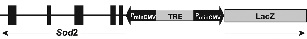
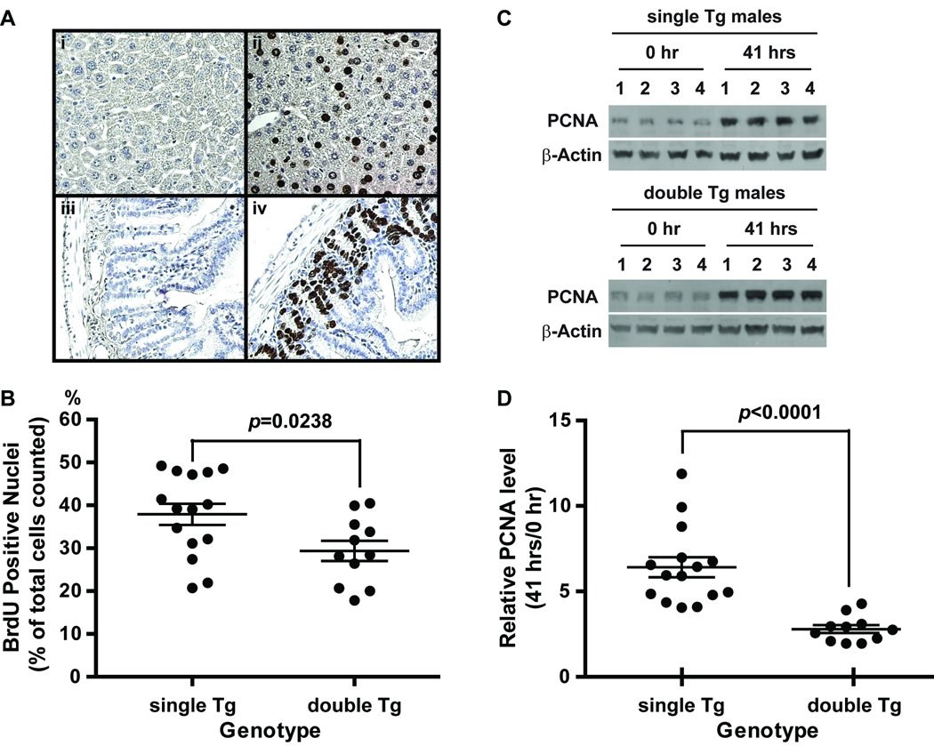
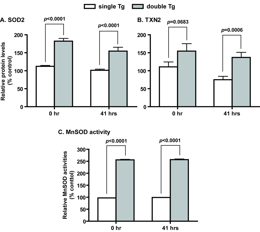
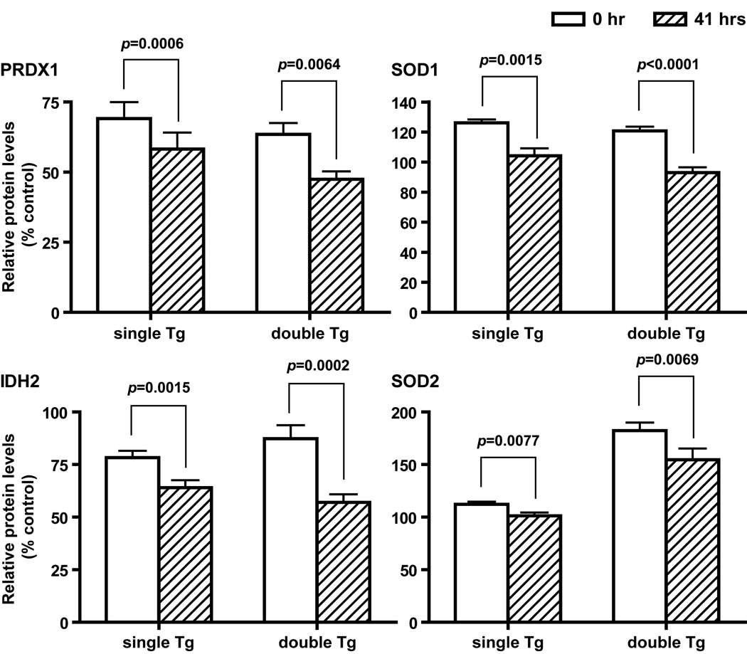
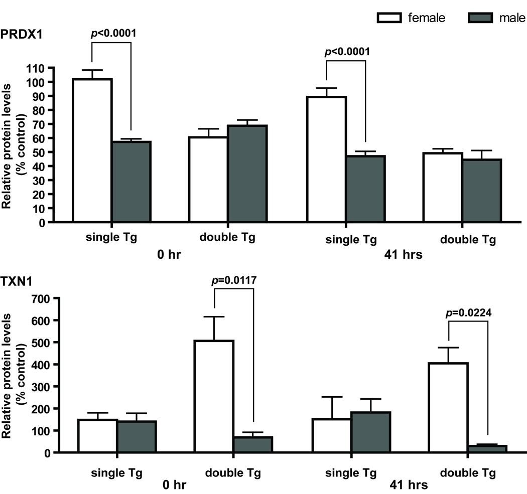
Mn superoxide dismutase (MnSOD) is an important mitochondrial antioxidant enzyme, and elevated MnSOD levels have been shown to reduce tumor growth in part by suppressing cell proliferation. Studies with fibroblasts have shown that increased MnSOD expression prolongs cell cycle transition time in G1/S and favors entrance into the quiescent state. To determine if the same effect occurs during tissue regeneration in vivo, we used a transgenic mouse system with liver-specific MnSOD expression and a partial hepatectomy paradigm to induce synchronized in vivo cell proliferation during liver regeneration. We show in this experimental system that a 2.6-fold increase in MnSOD activity leads to delayed entry into S phase, as measured by reduction in bromodeoxyuridine (BrdU) incorporation and decreased expression of proliferative cell nuclear antigen (PCNA). Thus, compared to control mice with baseline MnSOD levels, transgenic mice with increased MnSOD expression in the liver have 23% fewer BrdU-positive cells and a marked attenuation of PCNA expression. The increase in MnSOD activity also leads to an increase in the mitochondrial form of thioredoxin (thioredoxin 2), but not in several other peroxidases examined, suggesting the importance of thioredoxin 2 in maintaining redox balance in mitochondria with elevated levels of MnSOD.


Figures

Figure 1
Figure 1
Inducible MnSOD (Sod2) expression construct under the control of a bi-directional tet-responsive element (TRE). The thick vertical bars in Sod2 represent the five exons and the horizontal line represents the intron sequence. The arrows indicate the orientations of Sod2 and LacZ. The drawing is not to the scale.
Figure 2
Figure 2
BrdU and PCNA analysis of liver samples from partial hepatectomy. A, Images of BrdU staining in liver and small intestine recovered at 41 hours post surgery. Representative BrdU positive sections (ii and iv) and negative controls (i and iiI) are shown. BrdU positive cells showed intense brown color in the nuclei interspersed among hepatocytes (ii) and at the base of the crypts of the small intestine lining (iv). Sections were counter stained with hematoxylin. Small intestines were used as positive controls for BrdU injection and immunostaining. Negative controls (i and iii) for BrdU immunostaining were carried out without the primary antibody. B, Percentage of BrdU positive cells in livers recovered at 41 hours post surgery from single and double Tg mice. C, PCNA levels determined by western blot analysis. Representative results from 4 each independent pairs of liver samples collected at 0 hour and 41 hours post surgery from male single and double Tg mice are shown. D, the ratio of PCNA levels between 0 hour and 41 hours. Single Tg, Sod2-TRE-LacZ single transgenic mice; double Tg, Sod2-TRE-LacZ/LAP-tTA double Tg mice. Single Tg, n=15 (11 males and 4 females); double Tg, n=11 (4 males and 7 females). Scatter plots show individual values and horizontal lines indicate mean ± SEM in B and D.
Figure 3
Figure 3
Increased expression of MnSOD and TXN2 in Sod2-TRE-LacZ/LAP-tTA double Tg mice. A and B, MnSOD and TXN2 protein levels determined by western blot analyses. Single Tg, n=15 (11 males and 4 females); double Tg, n=11 (4 males and 7 females). C, MnSOD activities determined by non-denaturing IEF gel electrophoresis followed by activity staining. Single Tg, n=8 (4 males and 4 females); double Tg, n=11 (4 males and 7 females). SOD2 and TXN2 are protein designations for MnSOD and thioredoxin 2, respectively. Mean ± SEM of normalized signal intensity are shown. Error bars are too small to show in the activity figure. Student’s t test was carried out for the comparison between single and double Tg at each time point.
Figure 4
Figure 4
Changes in cytosolic and mitochondrial aconitase activities. ACO1 and ACO2 were separated by cellulose acetate gel electrophoresis followed by activity staining. Single Tg, n=8 (4 males and 4 females); double Tg, n=11 (4 males and 7 females). ACO1 and ACO2 are protein designations for cytosolic aconitase and mitochondrial aconitase, respectively. Mean ± SEM of normalized signal intensity are shown.
Figure 5
Figure 5
Decreased antioxidant protein levels after partial hepatectomy. Proteins that show a significant reduction in both single and double Tg mice at 41 hours post surgery are shown. Mean ± SEM of normalized western blot signals are shown. Single Tg, n=15 (11 males and 4 females); double Tg, n=11 (4 males and 7 females). PRDX1, SOD1, IHD2, and SOD2 are protein designations for peroxiredoxin 1, CuZnSOD, isocitrate dehydrogenase 2 (NADP-dependent), and MnSOD, respectively. Paired t test was carried out for the comparison between 0- and 41-hour samples within each genotype.
Figure 6
Figure 6
Gender and genotype interaction affects PRDX1 and TXN1 levels at both time points. Upper panel, significant differences in PRDX1 protein levels between males and females were observed in single Tg mice. At both 0 and 41 hours, male single Tg mice have less than 60% of PRDX1 found in female single Tg mice. No significant differences were observed between male and female double Tg mice. Lower panel, significant differences in TXN1 protein levels between males and females were observed in double Tg mice. TXN1 levels in male double Tg mice are only 15% and 8% of that in female double Tg mice, respectively. No significant difference in TXN1 level between males and females was observed in single Tg mice. Single Tg, n=15 (11 males and 4 females); double Tg, n=11 (4 males and 7 females). Two-way ANOVA was used to determine interaction between gender and genotype; Student’s t test was used for the comparison between males and females within each genotype.
Figure 7
Figure 7
Gender differences in GR and GST activities. Upper panel, significant differences in GR activities between males and females were observed in single Tg mice at both time points. GR activities in male single Tg mice were 40 and 60% higher than that in females at 0 and 41 hours, respectively. Lower panel, significant differences in GST activities between males and females were observed in single and double Tg mice at both 0 and 41 hours. On the average, GST activities in male mice were 80 to 110% higher than that in females. GR and GST are protein designations for glutathione reductase and glutathione S-transferase, respectively. Single Tg, n=14 (10 males and 4 females); double Tg, n=11 (4 males and 7 females). Mean ± SEM of specific activities are shown. Student’s t test was used for the comparison between males and females within each genotype.
Figure 8
Figure 8
Reduction of 3-nitrotyrosine in mouse livers after partial hepatectomy. Representative western blot results from four double Tg males are shown (upper panel). Signal intensities from the two major 3-nitrotyrosine positive bands were quantified and normalized to that of β-actin (lower panel). Sizes of the two major 3-nitrotyrosine positive bands were determined based on molecular weight standards and actual locations of IDH2 and MnSOD on the same blot. Paired t test was carried out for the comparison of each 3-nitrotyrosine positive band between 0- and 41-hour samples.

References

    1. Li Y, Huang TT, Carlson EJ, Melov S, Ursell PC, Olson JL, Noble LJ, Yoshimura MP, Berger C, Chan PH, Wallace DC, Epstein CJ. Dilated cardiomyopathy and neonatal lethality in mutant mice lacking manganese superoxide dismutase. Nat Genet. 1995;11:376–381. - PubMed
    1. Chen Z, Siu B, Ho YS, Vincent R, Chua CC, Hamdy RC, Chua BH. Overexpression of MnSOD protects against myocardial ischemia/reperfusion injury in transgenic mice. J Mol Cell Cardiol. 1998;30:2281–2289. - PubMed
    1. Klivenyi P, St Clair D, Wermer M, Yen HC, Oberley T, Yang L, Flint Beal M. Manganese superoxide dismutase overexpression attenuates MPTP toxicity. Neurobiol Dis. 1998;5:253–258. - PubMed
    1. Yen HC, Oberley TD, Gairola CG, Szweda LI, St Clair DK. Manganese superoxide dismutase protects mitochondrial complex I against adriamycin-induced cardiomyopathy in transgenic mice. Arch Biochem Biophys. 1999;362:59–66. - PubMed
    1. Epperly MW, Kagan VE, Sikora CA, Gretton JE, Defilippi SJ, Bar-Sagi D, Greenberger JS. Manganese superoxide dismutase-plasmid/liposome (MnSOD-PL) administration protects mice from esophagitis associated with fractionated radiation. Int J Cancer. 2001;96:221–231. - PubMed

Publication types

MeSH terms

LinkOut - more resources